Posted on

FLUSHING PARASITES FROM YOUR BODY NATURALLY!

As we have been talking about cleansing the liver and gall bladder in the last couple of issues, I thought that I would continue the same thread and talk a little about cleaning out parasites from all body organs and tissues. “I don’t have any of those,” I hear you saying. Well, think again, and look at the statistics.

PARASITES EVERYWHERE
According to the publication in 1995 “Parasitic Diseases” it is reported:

Nematodes (Round Worms) – 1 billion individuals
Cestodes (Tape Worms) – 300 million individuals
Tremadodes (Flukes) – 300 million individuals
Protozoa (Amoebas) – 1 billion individuals
Arthropods (Insects para) – 500 million individuals

It is clear from this data that parasites are not confined only to third world countries! An interesting report by the Parasitologist, Omar Amin, Ph.D. states that in a five star restaurant, samples were taken from under the fingernails of the entire staff and 100% of them tested positive for feces under their nails. “Bon Appetit!”

We are always picking up parasites! The main source is undercooked meats, followed by household pets. After we are infected we can give it to each other via blood (childbearing), saliva (kissing), semen (lovemaking) and breast milk (nursing).

Dr. Hulda Clark in her book “The Cure For All Cancers” says that the human intestinal fluke Fasciolopsis buskii is the cause of most cancers. When these reach the liver, a growth factor is produced – ortho-phospho-tyrosine – that make cells divide and cancer proliferates.

Orthodox medicine tends to use a variety of drugs (with bad side-effects) to kill specific parasites, but this is not very practical when there are many different varieties present. Dr. Clark usually finds on average a dozen or more parasites in each patient, out of the 120 that she tests for. Dr. Clark has found three herbs that can kill ALL these parasites in the body without the dreadful side-effects of drugs. Below is a modified version of her treatment:

PARASITE KILLING PROGRAMME
Three herbs used together can get rid of over 100 different parasites without so much as a headache or nausea. The herbs are black walnut tincture, wormwood, and common cloves. Black walnut hull and wormwood kill adults and developmental stages of at least 100 parasites. Cloves kill the eggs. They must be used together as a single treatment for them to be effective. The detailed program is as follows:

1. Black Walnut Hull Tincture – take in water or juice before meals on an empty stomach

Day 1 – Day 6 – take 2 teaspoons of the Walnut tincture in 1/2 cup water a little before meals, once daily.
For maintenance – continue to take 2 teaspoons only once weekly for about 3 months.

2. Wormwood Combination Capsules: – take these before dinner with water or juice

Day 1 – 1 capsule
Day 2 – 1 capsules
Day 3 – 2 capsules
Day 4 – 2 capsules
Day 5 – 3 capsules
Day 6 – 3 capsules
Day 7 – 4 capsules
Day 8 – 4 capsules
Day 9 – 5 capsules
Day 10 – 5 capsules
Day 11 – 6 capsules
Day 12 – 6 capsules
Day 13 – 7 capsules
Day 14 – 7 capsules

Take all the capsules together as a single dose. As a maintenance dose take 7 capsules once a week for 3 months.

3. Cloves Capsules – take before meals.

Day 1 – Take one capsule 3 times daily before food.
Day 2 – Take two capsules 3 times daily before food.
Days 3 to 10 Take 3 capsules three times daily.

After day 10, take 3 capsules once daily for 3 months.

MAINTENANCE PROGRAMME
These parasites are being picked up all the time, that’s why it’s important to stay on a maintenance programme, once weekly, as follows:

Black Walnut Tincture – 2 tsp. before eating once weekly
Wormwood capsules – 7 capsules once weekly before eating
Cloves – 3 capsules once a day on an empty stomach.

You may take these at different times of the day, or all together.

It is highly recommended that everyone go on a parasite cleanse at some point or other. Certainly in clinical practice I have seen amazing recoveries from chronic problems. One such case was a South African lady who recovered in record time – about 2 months – from a 20-year old chronic fatigue syndrome just after completing her parasite cleanse! There are other similar cases that I have recorded in my clinical archives. I have been through the cleanse myself too, and it is true that you develop minimal symptoms. I felt a little “spacey” the first day, which may have been due to a “die-off” reaction.

Good luck with the cleanse. If you have any interesting experiences, let me know!

The Food and Drug Administration have not evaluated these statements. This information and products are not intended to diagnose, treat, cure or prevent any disease.

by Dr. George J Georgiou, Ph.D.,N.D.,D.Sc (A.M)
Natural Medicine Practitioner
drgeorge@avacom.net
www.naturaltherapycenter.com

Posted on 116 Comments

FLUSH GALL STONES NATURALLY!

Apple Juice image

FLUSH GALL STONES NATURALLY!

I have personally supervised more than 200 people in clinical practice who have been through the gall bladder flush described below. I have never encountered problems more than the occasional nausea, assuming that the protocol is followed as mentioned below.

HUNDREDS OF STONES
It is estimated that 20% of the world’s population will develop gallstones in the gall bladder at some stage in their lives. This figure does not take account of the numerous stones that accumulate in the liver and its ducts, given that the liver produces them in the first place, and then travel down to the gall bladder.

I have personally witnessed the removal of gallstones from hundreds of patients – some of them had gall-bladder symptoms but had no positive results from an ultra sound scan. Most, however, did not have any symptoms at all, yet would flush out literally hundreds of stones – no exaggeration! One woman in her 50’s had three scans and the radiologists found nothing. She had pains in the gall-bladder region for 20 years. When she did the gall bladder flush she removed 430 stones the first time around, and about 300 the second time!

About a week before I did my first gall bladder flush I went to see a friend who is an ultrasound specialist. He checked my gall bladder and found it as clean as a whistle. When I flushed a week later I removed 5 LARGE stones (about the size of a hazelnut), and about 150 smaller stones, including gravel.

It is believed by many naturopathic doctors that EVERYONE has gallstones, some less than others, and I have validated this many times in clinical practice.

The cleanse that I recommend below takes place within a period of less than 14 hours and can be done at home over the weekend. It is a painless and harmless natural way of removing stones, without requiring invasive procedures such as surgery, laser, etc.

GALL BLADDER CLEANSE PROTOCOL 

VERY IMPORTANT: It is crucially important BEFORE you begin this gall bladder cleanse to have drunk TWO GLASSES of APPLE JUICE (packaged juice is just as good as fresh) every day for 14 days before the cleanse. This is very important as certain constituents of apple juice (it is believed to be the high pectin levels) SOFTEN the stones and enable them to pass HARMLESSLEY through the gall ducts.

I have supervised hundreds of such cleanses using exactly this protocol that I am recommending here without one patient suffering any harm whatsoever. But please follow the instructions carefully, and it must be said that you have ultimate responsibility, given that none of you are actual patients of mine.

An alternative to apple juice that is just as effective, is Orthophosphoric acid (75%) – take 30 drops a day for 3-4 days, and gradually increase the dosage to 50 drops daily – continue for 10 days. Each 30 drops contain 390mg of Orthophosphoric acid.

Take no medications, vitamins or pills that you can do without on the day of the cleanse. They could prevent success.

Eat a NO-FAT breakfast and lunch such as cooked cereal with fruit, fruit juice, brown bread with a little honey (no butter, milk or margarine), baked potato or other vegetables with salt only.

2.00 PM Do NOT drink or eat after 2 o’ clock.

6.00 PM Drink one serving (3/4 cup) of ice cold Epsom salts. Mixing one-tablespoon Epsom salts into 3/4 cup cold water can make this. You may add 1/8 teaspoon of vitamin C powder to improve the taste. You may drink a little water afterwards, or rinse your mouth out.

8.00 PM Repeat the Epsom salt drink as above.

9.45 PM Pour 1/2 cup olive oil and squeeze 1/2 cup orange or grapefruit juice into this, with one juice of a whole fresh lemon. Shake or stir hard until the oil and fruit juice mix thoroughly.

Visit the bathroom now, as you will lie down as soon as you take the olive oil mixture.

10.00 PM Drink the olive oil and juice you have mixed. Drinking through a plastic straw helps it go down easier. Drink it standing up, not sitting or lying. You may use a little honey between sips to help it down. Try to drink it as quickly as you can, within 5 minutes.

LIE DOWN IMMEDIATELY, ON YOUR RIGHT SIDE! You may fail to get stones out if you don’t. The sooner you lie down, the more stones you will get out. Try to keep perfectly still for 20 minutes. You may feel a train of stones traveling along the bile ducts like marbles. There is no pain because the bile duct valves are open, thanks to the Epsom salts. GO TO SLEEP.

NEXT MORNING – upon awakening take another dose of Epsom salts. Drink 3/4 cup of the mixture. You may go back to bed. Don’t drink this before 6.00 a.m.

2 HOURS LATER – take you 4th and last dose of Epsom salts. Drink 3/4 cup. You may again go back to bed.

AFTER 2 MORE HOURS – You may eat. Start with fruit juice. Half an hour later eat fruit. One hour later you may eat regular food but keep it light – salads, steamed vegetables, fruit, juices, etc. By supper you should feel well. There are occasions when you may feel a little unwell for a couple of days, particularly when you have not done a liver flush before the gall bladder cleanse. Sometimes this may be due to stones and debris remaining in the colon and causing irritation and inflammation. Colon hydrotherapy or a good, deep enema can help this problem.

In the morning expect diarrhoea. Try to catch the gallstones in a sieve in the toilet pan so that you can see them. If any of you have a camera please take photos and send me a copy for my clinical archives. Most of the stones will be SOFT and green, breaking easily, or even dissolving. All these green stones are as soft as putty thanks to the apple juice, and are mostly made of cholesterol.

A few days after the cleanse, stones from the rear of the liver will have traveled “forward” towards the main bile ducts leaving the liver, and fill the gall bladder again! This is why it is sometimes necessary to do up to 6 cleanses (perhaps one each month) in order to get rid of all the stones. If a cleanse produces no more stones, your liver can be considered to be in excellent condition!

Wish you all well! A clean gall bladder can lead to higher energy levels, alleviation of allergies, flatulence, digestive problems, back pains and more.

The Food and Drug Administration have not evaluated these statements. This information and products are not intended to diagnose, treat, cure or prevent any disease.

by Dr. George J Georgiou, Ph.D.,N.D.,D.Sc (A.M)
Natural Medicine Practitioner

Posted on

OXYGEN FREE RADICALS AND AGING: PART II

Aging Image

Oxygen Free Radicals and Aging: Part II

by Emmanuel C. Opara, Ph.D.

In case you missed Part I of this three-part series, we reviewed the major theories of aging, including: the rate of living theory; the somatic mutation theory; the neuroendocrine theory; the glycation theory; the crosslinking theory; and the free radical theory. The free radical theory of aging, which holds that aging is due to damage to DNA and molecular membranes by highly reactive molecules known as free radicals, is especially appealing for many reasons. First, it provides a unifying mechanism for the different theories; second, it can be experimentally verified; third, it suggests a number of parameters that can be evaluated to determine the progress of aging and the success of anti-aging therapies; and finally, it is a mechanism that can be altered by appropriate antioxidant therapies. Sources of free radicals were also discussed, which included both exogenous (outside the body) and endogenous (inside the body) sources. Some of these exogenous sources include air pollution and cigarette smoke. It’s obvious that it’s impossible to avoid contact with these reactive molecules.

Fortunately, the body is naturally equipped with antioxidant defense systems to detoxify these dangerous agents. Unfortunately, the body’s defense system becomes less effective as we get older, leading to the accumulation of oxidative damage and the development of chronic degenerative diseases like arthritis, hypertension, atherosclerosis, diabetes, cancer and diseases of the central nervous system such as stroke, Alzheimer’s disease and Parkinson’s disease. Depletion of antioxidants can thus lead to a variety of chronic diseases. The relationship between oxidative damage and aging is a double-edged sword. On one hand, oxidative damage to DNA, lipids, proteins and other macromolecules appears to be a major contributing factor to aging,1 while at the same time, this oxidative damage accumulates with aging, despite attempts by an individual’s cellular machinery to repair it.7,8

For example, blood levels of glutathione, the major cellular antioxidant in the body, decline with age.1,2 Low glutathione levels are associated with a higher incidence of diseases in the elderly.3 In fact, blood glutathione concentrations may become recognized not only as a predictor of susceptibility to disease, but also as a marker of biological age.

Oxidation and Disease Progression
In this article, we’re going to discuss the specific contributions that damaging free radical reactions make in promoting the onset and progression of the chronic degenerative diseases of aging. We’ll discuss the role of free radicals (oxidants) in the development of each of these diseases, and provide a strong rationale for using antioxidant supplements for disease prevention and treatment. Indeed, a great deal of evidence regarding the role of free radicals in aging and its related diseases has been derived from studies in which antioxidant supplements have been used to successfully treat or prevent these diseases. Many of these studies will be reviewed in Part III of this series.

Cancer
Free radicals are involved in both the process of aging and the development of cancer.4 They attack many cellular targets including membranes, proteins and nucleic acids,5 and cause structural damage to the cellular DNA. These structural changes manifest as point mutations and chromosomal alterations in cancer-related genes.5 Consequently, elderly people are predisposed to the development of cancer. Fortunately, certain antioxidant supplements like vitamins C and E, can prevent much oxidative damage to DNA and thus reduce the ability of the oxidants to induce cancer.7

Cardiovascular Disease
One of the most significant discoveries in modern cardiovascular disease research was the discovery of how free radical activity contributes to cardiovascular disease.4,7 These discoveries were made using two different approaches. One approach, using epidemiological studies, showed that cardiovascular disease is associated with low plasma concentrations of antioxidant vitamins.7 The other approach, using experimental studies, provided strong evidence that free radicals oxidize low density lipoprotein (LDL) – the bad form of cholesterol. The modified LDL molecules are then easily taken up by white blood cells called macrophages (phagocytes) to form foam cells and plaques in the cardiovascular wall,4,7 causing a hardening and narrowing of the blood vessels which impairs blood flow and deprives the heart of oxygen and nutrients. Also, what is known as reperfusion (reoxygenation) injury is caused by inadequate supplies of intracellular antioxidants! Reperfusion injury is the damage to cells which occurs following restoration of the blood and oxygen supply to the heart after a period of ischemia (inadequate blood supply). Antioxidants are able to prevent or reduce the severity of this type of tissue damage.4

Diseases of the Central Nervous System
Oxidative damage has been implicated in brain aging as well as several degenerative diseases of the central nervous system (CNS). A number of mechanisms have been identified which explain the causative role played by free radicals in the neurodegenerative diseases. First, oxidants may activate an enzyme of nerve cells (poly ADP-ribose synthetase) and cause an increase in the intracellular calcium ion concentrations, which is injurious to neurons.12 Second, reoxygenation of brain cells following a period of ischemia contributes to oxidant injury of the CNS, as already mentioned in the case of cardiovascular disease. Third, certain areas of the brain, e.g., the globus, pallidus and substantia nigra often contain high amounts of iron. Excess iron enhances oxidative reactions and consequent injury to the neurons.12 It is therefore not surprising that antioxidant supplements have been successfully used to improve the functioning of the brain both in people who are aging normally, as well as patients suffering from neurodegenerative diseases such as stroke and Alzheimer’s and Parkinson’s diseases.

I. Strokes
Free radical-produced damage due to ischemia-reperfusion in the brain has been implicated as a major cause of strokes.12 Antioxidant supplements appear to be of benefit in the prevention and reduction of strokes.12,13 And, to top it off, recent clinical studies indicate that surgical therapy (i.e., carotid endarterectory) is actually worse than no therapy in many cases.

II. Alzheimer’s Disease
Alzheimer’s disease (AD) is the tragic brain-robbing disease of aging. It can be recognized by the progressive loss of memory and other aspects of cognitive functioning. Its characteristic pathological features include tangles of nerve fibers, senile plaques (which contain aluminum, iron, and calcium) and the loss of brain cells.9 Oxidative damage has been implicated in AD, for a number of reasons. First, the brain has the highest oxygen consumption rate of any organ in the body, high concentrations of easily-oxidizable lipids, and a relative deficiency of antioxidant enzymes (compared to other tissues).9 Second, iron, which plays an important role in free radical generation, has been found in high concentrations in the brain in AD.9 Third, antioxidant supplementation often improves memory performance in aged individuals.10

III. Parkinson’s Disease
The hallmark of Parkinson’s disease is degeneration of a portion of the brain called the substantia nigra – the portion of the brain that produces the stimulatory neurotransmitters epinephrine (adrenaline) and norepinephrine (noradrenaline). There are two mechanisms that have been proposed as possible causes of this neuronal damage.11 The first is that increased production of oxidants causes damage to this vital portion of the brain (the substantia nigra) through iron-dependent free radical reactions. It has been shown that the iron content of the substantia nigra is often elevated, while ferritin, the iron-binding protein is often decreased in Parkinson’s disease. Again, the resulting increase in free iron ions enhances iron-dependent lipid peroxidation and damage to nerve cells.12 A second mechanism is impaired neuronal mitochondrial function, which leads to failure of energy production and adverse changes in the metabolism of nerve cells.

There are several mutually supporting links between these two mechanisms.11 As previously noted, mitochondria are a critical target for damage by free radicals.4,7,11 Also, mitochondrial damage may cause a further increase in generation of free radicals.11 Another link between the two mechanisms (i.e., oxidative damage to mitochondria vs. free-radical induced damage to neuronal metabolism) is glutathione. Patients with Parkinson’s disease tend to have low levels of this powerful intra-cellular antioxidant in their brains, resulting in higher levels of free radical activity.11 These two mechanisms both contribute to neuronal damage through alterations in glutathione levels.11

A variety of antioxidant vitamins and drugs are helpful for patients with this disease, especially Vitamin E and deprenyl I believe that one of the most beneficial nutrients for Parkinson’s disease is N-acetyl cysteine (NAC). NAC boosts glutathione production, and should be of tremendous benefit to Parkinson’s patients, in doses ranging from 600-1200 mg per day.

Inflammatory Diseases
Arthritis and inflammatory diseases of the large intestine, such as ulcerative colitis and Crohn’s disease, are conditions in which oxidative damage has also been implicated.4,14 While there is agreement that free radicals play a role in the pathogenesis of inflammatory disease, the mechanism(s) involved is still a matter of intense research. One plausible mechanism of oxidant mediated injury involves tissue protein oxidation, otherwise referred to as protein carbonylation. It has been proposed that inflammatory bowel disease (IBD) may arise from the oxidation of proteins in the intestinal mucosa cells which thereby disrupt the critical enzyme systems that are important for the maintenance of mucosal integrity or ion transport, both of which are impaired in IBD.15 Antioxidant supplements have helped many people who suffer from a variety of inflammatory diseases.4,7,15

Diabetes
Diabetes is a multi-systemic disease caused by a defect in glucose metabolism. Abnormally high blood sugar levels are a major clinical feature of the disease, which is usually followed by accelerated onset and progression of atherosclerosis and other diseases. In type 2 diabetes, oxidative damage has been implicated in both the development of the disease as well as its many complications. Oxidants inhibit glucose metabolism in the glycolytic pathway and at the level of oxidative phosphorylation, thereby causing a sugar overload in the blood. The medical term for high blood sugar levels is hyperglycemia. Increased blood sugar levels cause auto-oxidation of glucose and glycation of proteins which have been implicated in the development of diabetic complications.16 It is therefore not surprising that antioxidant supplements – particularly vitamins C and E, and lipoic acid – have been found to be beneficial to patients suffering from diabetes.16 This subject was covered extensively in previous reviews by this author (see Nutritional News volume 10: 6 and 7, 1997).

Vision

I. Cataracts
Oxidative damage to the lens of the eye, which collects and focuses light on the retina, plays a major role in the formation of cataracts.7,17 With age, the constituents of the lens are damaged by oxidants causing opaque precipitations that are referred to as senile cataracts.17 A variety of antioxidant supplements have been shown to provide protection against the development of cataracts7,17 – especially, lipoic acid – a key player in the prevention of cataracts.

II. Macular Degeneration
Macular degeneration is a leading cause of blindness in the elderly. It is one condition which ophthalmologists used to stand by and helplessly watch as their patients’ vision failed. Now, they are finding that combinations of antioxidant and carotenoids have demonstrated the ability to halt or delay the progression of this dreaded condition.
Many studies have been performed with multi-nutrient protocols to treat ARMD and cataracts. For example, Dr. E.N. Crary of Smyrna, Georgia published a study in 1987 in the Southern Medical Journal titled “Antioxidant Treatment of Macular Degeneration of the Aging and Macular Edema in Diabetic Retinopathy.” In this study, Dr. Crary used 250 mcg of selenium, 15,000 units of beta carotene, 500 mg of vitamin C and 400 units of vitamin E for a period of 7 to 12 years. He treated over 500 patients and found that the treatments either halted or improved degenerative macular changes in 60% of the patients with age-related macular degeneration.

Summary
It is apparent, as outlined above, that free radical (oxidative) damage plays a significant role in the process of aging as well as in the development of a broad spectrum of age-associated diseases. Specific supplements and their dosages for preventing and minimizing the effects of these diseases will be reviewed in part III of this series.

References:

1. Lang CA, Naryshkin S, Schneider DL, Mills BJ, Lindeman RD. Low blood glutathione levels in healthy aging adults. J Lab Clin Med 120: 720-725, 1992.
2. Vina J, Sastre J, Anton V, Bruseghini L, Esteras A, Aseni M. Effect of Aging on glutathione metabolism. Protection by antioxidants. In: Free Radicals and Aging. Emerit I and Chance B (eds.), 136-144, 1992.
3. Julius M, Lang CA, Gleiberman L, Harburg E, DiFranceisco W, Schork A. Glutathione and morbidity in a community-based sample of elderly. J Clin Epidemiol 47: 1021-1026, 1994.
4. Cross CE, Halliwell B, Borish ET, Pryor WA, Ames BN, Saul RL, McCord JM, Harman D. Oxygen Radicals and Human Disease. Ann Int Med 107: 526-545, 1987.
5. Cerutti P, Ghosh R, Oya Y, Amstad P. The role of the cellular antioxidant defense in oxidant carcinogenesis. Environmental Health Perspectives 102 (suppl 10): 123-130, 1994.
6. Block G. A role for antioxidants in reducing cancer risk. Nutrition Reviews 50: 207-213, 1992.
7. Ames BN, Shingenaga MK, Hagen TM. Oxidants, antioxidants and the degenerative diseases of aging. Proc Natl Acad Sci (USA) 90: 7915-7922, 1993.
8. Gilchrist BA, Bohr VA. Aging processes, DNA damage, and repair. FASEB J 11: 322- 330, 1997.
9. Lovell MA, Ehmann WD, Butler SM, Markesbery WR. Elevated thiobarbituric acid- reactive substances and antioxidant enzyme activity in the brain in Alzheimer’s disease. Neurology 45: 1594-1601, 1995.
10. Perrig WJ, Perrig P, Stahelin H. The relation between antioxidants and memory performance in the old and very old. J Am Geriatr Soc 45: 718-724, 1997.
11. Di Monte DA, Chan P, Sandy MS. Glutathione in Parkinson’s disease: a link between oxidative stress and mitochondrial damage. Ann Neurol 32: S111-S115, 1992.
12. Halliwell B. Oxidants and the central nervous system: some fundamental questions. Acta Neurol Scand 126: 23-33, 1989.
13. Mobarhan S. Micronutrient supplementation trials and the reduction of cancer and cerebrovascular incidence and mortality. Nutrition Reviews 52: 102-105, 1994.
14. Ruan EA, Simbasiva R, Burdick S, Stryker SJ, Telford GL, Otterson MF, Opara EC, Koch TR. Glutathione levels in chronic inflammatory disorders of the human colon. Nutrition Research 17: 463-473, 1997.
15. Otamiri T, Sjodahl R. Oxygen radicals: their role in selected gastrointestinal disorders. Dig Dis 9: 133-141, 1991.
16. Paolisso G, Giugliano D. Oxidative stress and insulin action: is there a relationship? Diabetologia 39: 357-363, 1996.
17. Taylor A. Effect of photooxidation on the eye lens and role of nutrients in delaying cataract. In: Free Radicals and Aging. Emerit I and Chance B (eds.), 266-279, 1992.

Posted on

NON-DRUG SUPPORT FOR BREAST AND PROSTATE CANCERS

pink ribbon for breast cancer iamge

An Interview with Shari Lieberman, PhD, CNS, FACN

by Jim English

Last month, in Part I of our interview with noted researcher, scientist and author, Shari Lieberman, Ph.D., CNS, FACN, we discussed her new Auto Immune Formula, which is designed to support, modulate and enhance healthy immune function. In Part II we continue our interview with Dr. Lieberman, discussing the development of two new cancer-support formulas, BCF (Breast Care Formula) and PCF (Prostate Care Formula).

VRP: Thank you for meeting with us again, Dr. Lieberman. Last month we were discussing your Auto Immune Formula, but you have also recently introduced BCF (Breast Care Formula) and PCF (Prostate Care Formula). Can you describe these formulas and how they work?

Dr. Lieberman: While the Auto Immune Formula was designed to support, stimulate and enhance healthy immune function, my aim in designing the Breast and Prostate Care Formulas was to address the needs of people either diagnosed with, or concerned about cancers of the breast or prostate. While these are, obviously, different diseases, they share important similarities that research shows can benefit from factors common to both formulas.

Specifically, I wanted to address a common problem encountered by cancer patients. Many cancer patients spend a great deal of time seeking out and researching the medical literature relating to their diseases. Ultimately they come across the large body of published research and clinical data that reveals how antioxidants and other natural compounds can either directly support recovery from their disease, or protect them from the terrible side effects of conventional anticancer therapies. And once they discover this information, it is only natural that they want to incorporate these compounds with their therapy.

Now the problem is that when a patient tells their oncologists that they want to start taking antioxidants to help them deal with the side effects of chemotherapy and radiation therapy, their oncologist usually objects. By training, cancer specialists will not allow their patients to use any compound that they’re not familiar with, and this leaves the patients in a quandary.

To get around this problem I decided to work with an oncologist who was versed in the medical research on natural therapies. Specifically I was looking for a cancer specialist who could not only verify the benefits of these compounds based on peer-reviewed studies, but ideally someone who was experienced in the use of these natural agents in their own clinical practice.

VRP: So you were looking for an oncologist who was familiar with the research and who also had clinical experience in using these ingredients to help patients with cancer. Was it your hope that in this way you would be able to help the patient to overcome the objections of traditional oncologists?

Dr. Lieberman: Exactly. If these formulas were going to be of value I really wanted to overcome the most common objections by getting input from an oncologist who had experience in using this approach, someone who could verify the safety and effectiveness of these compounds for treating cancer.

VRP: And eventually you collaborated with Dr. Douwes?

Dr. Lieberman: Yes. Friedrich Douwes, MD, is a noted oncologist and the Executive Medical Director of the St. George Hospital Cancer Treatment Center in Bad Aibling, Germany. I had visited his clinic several times and was aware of his tremendous experience in using these compounds with many patients. I knew that Dr. Douwes was very familiar with using natural therapies along with conventional treatments, and that he had reviewed the literature and clinical data on alternative therapies. When we sat down to begin working on these formulas we outlined four guiding principals.

First, each ingredient had to be backed by published research. Second, the mechanism of a given compound’s actions had be understood. Third, the ingredients had to be completely non-toxic. And fourth, we would only include ingredients that would not interfere in any way with conventional anticancer therapies.

We set out to review the ingredients that we knew were helpful in cancer. Some of the ingredients were proven to help patients by protecting healthy cells while making chemotherapeutic drugs more selective for cancer. Other ingredients were shown to help by making chemotherapy in and of itself more effective.

VRP: So working together you were able to develop formulas that you knew could be used safely and effectively by cancer patients. So what happens when a patient goes to their oncologist and tells them they are using these formulas?

Dr. Lieberman: Our hope is that the patients won’t find themselves locking horns with their oncologists. The point of these formulas is that they have no downside. They were designed to be used alone, or concomitantly, along with all of the current anticancer therapies.

This is the main reason I felt it was imperative to develop these formulas with an experienced and highly regarded oncologist. Dr. Douwes is one of the few oncologists I’ve ever met who really knows about all of these natural compounds. And Dr. Douwes has been using natural compounds for over two decades. In my experience that is extremely rare for an oncologist. I have many colleagues doing anticancer therapy, but very few of them are oncologists.

VRP: Is that why both formulas contain nutrients, such as Coenzyme Q10 or Selenium?

Dr. Lieberman: Yes, exactly. In the case of selenium, epidemiological studies have consistently shown that low serum levels of selenium are directly linked to an increased risk of developing prostate cancer. In fact, selenium has been shown to block the growth cycle of cell reproduction and induce apoptosis (programmed cell death) in prostate cancer cells. And in women, selenium has been shown to inhibit VEGF (vascular endothelial growth factor) in breast cancer cells. Research also shows that selenium reduces the incidence of malignant cells in animal models, and enhances the effects of chemotherapeutic drugs, such as Taxol® and adriamycin.

In the case of Coenzyme Q10, cancer patients are often very seriously deficient in this nutrient. CoQ10 is an antioxidant that protects against numerous reactive oxygen species (ROS) that are involved in cancers, particularly breast cancer. Most impressively, researchers have reported cases of induced remission in late stage breast cancer patients taking CoQ10, and published reports indicate that it may also be therapeutic for patients with prostate cancer.

VRP: Quercetin, like Selenium and CoQ10 is another powerful antioxidant. Is that why you have quercetin in both formulas, or is it there for another purpose?

Dr. Lieberman: Well, the list of anti-cancer properties of quercetin is really impressive. First, quercetin is a plant phytochemical similar to, and in some ways, more powerful than vitamin C. And like vitamin C, quercetin works by multiple mechanisms to prevent and fight cancers.

First, quercetin is a potent aromatase inhibitor that prevents the conversion of DHEA and testosterone to estrogen, which is well established for its role in promoting both breast and prostate cancers. But that’s just the beginning. Quercetin also prevents cancer by inhibiting estrone sulfatase in the liver, stimulating the immune response, and by triggering cancer cell apoptosis. It also helps by inactivating potentially carcinogenic liver enzymes while inhibiting the proliferation of cancer metastases.

And in a recent paper, researchers found that quercetin inhibits the cancer-causing effects of hydrogen peroxide. Cells must communicate with each other to stay healthy and to promote normal cell growth. Hydrogen peroxide is a known tumor promoter that works, in part, by inhibiting normal cell communication. What researchers discovered from animal studies is that quercetin works to inhibit this tumor-promoting property of hydrogen peroxide. The research, just published at the beginning of the year, may help to explain why cancer rates are often lower in people who eat foods high in phytochemicals and vitamin C.

And if all of this weren’t enough, quercetin also works synergistically with numerous drugs used in the treatment of cancer, including ribavarin, adriamycin and tamoxifen.

VRP: Very impressive. Now, lycopene has been well researched as an anti-cancer nutrient, particularly for preventing prostate cancer, yet I see that you have it in both the Breast and Prostate cancer formulas. Why is that?

Dr. Lieberman: To begin with, lycopene is the most potent carotenoid inhibitor of breast and prostate cancer progression. Research has shown that lycopene interferes with insulin-like growth factors (IGF) that are potent promoters of breast and prostate cancer. By interfering with these growth factors lycopene may reduce both the occurrence and the progression of breast and prostate cancers.

For example, recent epidemiological studies show that lycopene reduces the risk of prostate cancer. And additional research suggests that lycopene may also decrease the growth and spread of prostate cancers. In a Phase II study of men undergoing radical prostatectomy (removal of prostate), 73 percent of men given supplemental lycopene before surgery showed no spread of the cancer beyond the prostate. And in the group of controls-men not receiving supplemental lycopene-these numbers were almost reversed, and in 82 percent of the men their cancer had spread.

Additionally, plasma prostate-specific antigen (PSA) levels dropped 18% in the lycopene group, while increasing 14% in control group. These results suggest another mechanism by which lycopene decreases growth of prostate cancers.

VRP: Two items caught my eye when going over the ingredients list – Lactobacillus sporogenes and Convolvulus arvensis. What can you tell me about these ingredients?

Dr. Lieberman: Well, besides the strange names, both of these ingredients are powerful anti-cancer nutrients. Lactobacillus sporogenes is-as the name implies-a naturally occurring bacteria that supports a healthy intestinal environment by re-establishing the ‘good’ flora in the gastrointestinal tract and preventing the overgrowth of pathogenic bacteria. And we know that people who consume yogurt with a good source of lactobacillus bacteria have a lower risk of developing breast cancer. Additionally, lactobacillus has been shown to reduce the reabsorption of estrogens, and it has an antiproliferative effect on cancer cells.

The second ingredient you mentioned, Convolvulus arvensis extract, is the source of a group of proteoglycan molecules that have been shown to inhibit angiogenesis, the process used by tumors to promote the growth of new blood vessels that are necessary for tumor growth. In animal models Convolvulus arvensis extracts have been shown to halt tumor growth, and in higher concentrations cause a significant reduction in tumor size. In some animal models the extract completely eradicated the tumors. Human studies are currently underway.

VRP: Let’s move on to something more people may be familiar with – mushrooms. The last ingredient common to both formulas is an extract from the Maitake mushroom. What can you tell us about this extract?

Dr. Lieberman: Breast and prostate cancers benefit from Maitake D-Fraction, which has been shown to activate the immune system to kill cancer cells without harming healthy ones. Maitake activates cytotoxic T-cells that mark cancer cells for destruction, as well as the NK (Natural Killer) cells that actively seek out and destroy tumor cells. Maitake D-Fraction also increases the production of interleukin-1 and induces apoptosis of cancer cells.

Another important property of Maitake is that, if given concomitantly with either chemotherapy or radiation therapy, it enhances the effectiveness of these treatments while also reducing their unpleasant side effects. And it does this without reducing or hampering the effectiveness of either therapy.

Breast Care Formula

VRP: We’ve covered ingredients in both formulas. Let’s begin with the Breast Cancer Formula, which contains the medicinal mushroom, Shiitake.

Dr. Lieberman: Shiitake is a Chinese herb that is used in Japan as a powerful anti-cancer treatment. In addition to stimulating and enhancing the responsiveness of the immune system, shiitake has also been found to shrink tumors and prevent the spread of cancers. Shiitake is also synergistic with other chemotherapeutic drugs and has been found to prevent the recurrence of breast cancer after surgery. Shiitake has been shown to significantly prolong the lifespan of patients with breast and other cancers.

Another compound included in the Breast Cancer Formula is D-limonene. This unique compound contains an active ingredient known as perillyl alcohol that inhibits the growth and cell cycle progression of breast cancer cells.

VRP: You’ve also included DIM (diindolylmethane) which has been in several previous newsletter articles, primarily for its estrogen balancing and anticancer properties.

Dr. Lieberman: Yes, indole-3-carbinol (I3C) has been shown in both Phase 1 and Phase 2 human studies to be effective in inhibiting breast cancer growth in both estrogen positive and negative breast cancer cells. DIM is the active plant compound in I3C. DIM is also synergistic with chemotherapeutic agents such as tamoxifen. As a preventive supplement, DIM has been shown to lower breast cancer risks by reducing levels of 16-alpha-hydroxyestrone (a biological marker for breast cancer risk) while improving the ratio of 16-alpha-hydroxyestrone to 2-hydroxyestrone. By improving this ratio, DIM is effectively lowering the amount of ‘bad estrogen’ relative to the ‘good estrogen.’ Additionally, DIM modifies cytochrome p450 enzyme activity to support detoxification of carcinogens, induces apoptosis of breast cancer cells, and may stimulate progesterone production.

VRP: BCF also contains a good dose of Green Tea extract.

Dr. Lieberman: Yes, I’ve included Green Tea and Grape Seed extracts, both very powerful antioxidants that help to induce cell death in breast cancer cells. Additionally, Green Tea contains the active ingredient, epigallocatechin gallate, that has been shown in human studies to inhibit human breast cancer cell proliferation, reduce tumor invasion and metastasis, and prevent recurrence of breast cancer in early stage cases (stage I and II).

Prostate Care Formula

VRP: Moving on to the Prostate Care Formula, I see you’ve included several ingredients proven to help prevent prostate cancer.

Dr. Lieberman: Yes, all of these are well-established for their effects. Zinc, for example has been shown to inhibit prostate tumor cell invasion and metastasis while inducing apoptosis in prostate cancer cells.

I’ve also included Beta-sitosterol, which inhibits growth of prostate cancer, decreases cancer cell number and induces apoptosis. Animal studies support the use of beta-sitosterol in inhibiting prostate cancer growth, and epidemiological studies suggest beta-sitosterol is preventive for prostate cancer.

VRP: You also have Pygeum, also known for supporting prostate health.

Dr. Lieberman: Yes, Pygeum africanum is very effective for the treatment of benign prostatic hyperplasia, working by reducing prostate cell proliferation and inhibiting growth factors that are responsible for prostate overgrowth. And Saw Palmetto works well with Pygeum, since it is also effective for the treatment of human benign prostatic hyperplasia. Saw palmetto also induces apoptosis and necrosis in human prostate cancer cells, while inhibiting cancer cell invasion (metastases).

VRP: The last ingredient here is Stinging Nettle. Comment?

Dr. Lieberman: I’ve included Stinging nettle, which is very effective for the treatment of human benign prostatic hyperplasia. Stinging nettle works by reducing prostatic cell proliferation and inhibiting growth factors responsible for prostate overgrowth. And Stinging nettle acts synergistically when combined with Pygeum africanum.

VRP: Thank you for taking the time to discuss your work with us, and for sharing these sophisticated nutritional formulas that are long overdue in the fight against cancer.

Dr. Lieberman: Thank you, it has been my pleasure.

Posted on

NATURAL PROTECTION AND RECOVERY FROM STRESS

Stressing man image

Natural Protection and Recovery From Stress

by Ward Dean, MD

As the consequences of our current national trauma unfold in the days and months ahead, each of us must confront the anxiety and stress of facing an unpredictable future. Chronic anxiety and long-term post-traumatic stresses have a devastating effect on mental, psychological and physical health.

The Father of Stress

Eastern researchers have a long history of searching for solutions to stress-related problems. In Russia, after years of scientific investigation, Soviet scientists were inspired by the groundbreaking work of Dr. Hans Selye, a Canadian professor and leading pioneer in stress research. Internationally acknowledged as ‘the father of stress,’ Dr. Selye authored more than 1,700 scholarly papers and 39 books which had a profound influence on Soviet scientists doing stress research for the Russian military, sports and space programs.

General Adaptation Syndrome

Among the most memorable of Selye’s contributions was his concept of the General Adaptation Syndrome (GAS). First described in 1936, the GAS involves three progressive stages:

Alarm: The alarm reaction is the first stage, characterized by surprise and anxiety when exposed to a new situation. The body reacts by producing epinephrine and norepinephrine, the ‘flight or fight’ hormones. Additionally, the adrenal cortex is stimulated to produce additional cortisol and related hormones.

Resistance: In this stage, the body learns to efficiently cope with stress. Ideally, this stage continues until the stressful situation is resolved, leading to a rapid return to the resting state. Unfortunately, our capacity for resistance is limited and highly individualized (i.e. what is stimulating to one person may be devastating to another) and worsen with age. (Fig. 1). Just as a chain breaks at its weakest link, exhaustion of our adaptive capacity can result in stress-induced disease.

Exhaustion: In the presence of long-term exposure to the same stressor, we enter the third stage of GAS — exhaustion, characterized by the depletion of energy reserves and loss of adaptational ability, leading to fatigue or other symptoms or diseases.

This third stage is also sometimes referred to as adrenal maladaptation, or hyperadaptosis (Dilman and Dean, 1992). Chronically elevated cortisol levels can result in the signs and symptoms resembling Cushing’s Syndrome. (Fig. 2) Adrenal dysfunction may be manifested by (1) an excess or inadequacy of cortisol, DHEA, ACTH and/or CRF; (2) relative imbalances of these hormones and releasing factors, and (3) loss of sensitivity of the hypothalamus and pituitary to the normal, inhibiting effects of these hormones.

Symptoms of patients suffering from adrenal maladaptation syndrome include:

• Fatigue

• Nervousness

• Severe PMS

• Salt craving

• Depression

• Inability to concentrate

• Carbohydrate craving

• Allergies (hay fever, asthma)

• Anxiety

• Headache

• Alcohol intolerance

• Muscular pain and tenderness

• Joint pains and tenderness (arthritis)

• Weakness

• Poor memory

• Palpitation

• Abdominal discomfort

• Alternate diarrhea and constipation

• Obesity

• Poor wound healing

• Glucose intolerance

• Moon face

• Purple striae

• Loss of bone density

Dr. Nicholai Lazarev

Selye’s ideas of the General Adaptation Syndrome (GAS) were readily accepted by leading Russian researchers, including Dr. Nicholai Lazarev, a pioneer in the then-emerging fields of toxicology and preventive medicine. Shortly after graduating medical school Lazarev discovered that different industrial chemicals, even in mild concentrations and small dosages, cause similar alarm reactions. When exposure is prolonged, the body adapts by altering its physiological response (resistance). This adaptive reaction tends to gradually disturb homeostasis, which is damaging to health.

Lazarev found that Selye’s publication on stress confirmed his own findings. Selye proved that a variety of stressors — not just chemicals — can cause non-specific stress reactions. Selye’s work was so influential, in fact, that Lazarev changed the direction of his work. He began looking not only for substances that could improve humans’ general resistance to toxins, but could also correct the general adaptation reaction to all kinds of stressors, including both mental and physical stress.

WWII Research

From the beginning of the second World War, Lazarev’s efforts shifted to finding substances that could help Soviet soldiers overcome fatigue and improve their performance on the battlefield. With the soldiers as unknowing guinea pigs for amphetamines and other stimulants, Lazarev learned that many drugs were very effective in improving performance in response to great challenges in extreme situations for very short terms. However, he realized that the stimulants were harmful when used for prolonged periods. When WWII ended, Lazarev switched his focus to natural alternatives.

Traditional Herbs

Lazarev was especially intrigued by a group of herbs that ancient medical traditions referred to as ‘elite’ or ‘kingly.’ In traditional Chinese medicine these herbs were classified as effective for increasing physical and mental capacity, reducing fatigue, improving resistance to disease, and promoting life extension. In China, soldiers used these herbs before battle. In Siberia, hunters used the herbs before long and dangerous journeys. In 1948, Lazarev and his protege, Dr. Israel Brekhman, undertook the challenge of researching the utility and effectiveness of this group of plants that Lazarev named ‘adaptogens.’

The group first focused their efforts on ginseng, but quickly encountered problems with low availability, high cost, and wide fluctuations in effectiveness among different groups of subjects. This prompted Brekhman and his team to next focus their efforts on herbs adapted to growing in harsh environments based on the assumption that unique substances in these herbs enabled them to adapt and survive through millions of years of natural cataclysmic changes.

Eventually Brekhman’s group identified a group of herbs that fit the profile of an adaptogenic compound that is defined as: 1) an herb that is absolutely safe and possesses no side effects, even when used for long periods of time; 2) exhibits broad and nonspecific positive effects on the body; and 3) one that has a normalizing effect that keeps the body in balance while maintaining homeostasis and normalizing all functions.

Among the adaptogens discovered by Brekhman’s team are:

• Eleutherococcus (Siberian Ginseng)

Brekhman and his colleagues were particularly impressed with their studies of Eleutherococcus, commonly known as Siberian Ginseng, which they found had even greater anti-fatigue properties than the better-known Panax ginseng. Their studies soon led to a massive testing program with clinical trials conducted across the USSR on factory workers, long distance truck drivers, sailors on long voyages, and military personnel under severe stress. The stress studies revealed that Eleutherococcus helps to improve stamina, increases oxygen uptake, enhances physical energy and performance, and reduces high blood pressure.

Eleutherococcus was also found to enhance immune function, and incidence of cases during an influenza epidemic dropped by two-thirds. Additionally, 107 patients receiving anti-cancer drugs for gastric cancer showed evidence of a 50% reduction in impaired immunity, and a 50% decrease in drug dosage.

In 1962, Eleutherococcus, the prototype adaptogen, was declared an official herbal medicine by the USSR Minister of Health and was included in the National Drug Guide, the pharmacopoeia of the USSR. Eleutherococcus was soon widely available, and patients, athletes, cosmonauts, and soldiers used it regularly as a restorative. The Soviet government realized that this new class of natural remedy could give the Soviets an advantage in many areas, including sports, the arts, space, military, and medicine.

• Schisandra

Schisandra is an herb widely used in ancient and modern China to treat a number of conditions. It is a powerful antioxidant and anabolic agent. Schisandra also promotes glycogenesis, the process that converts carbohydrates into glycogen. Glycogen is stored in the liver and muscles until needed and then converted into glucose. This process helps spark the energy necessary to revitalize ‘stressed out’ cells.

Research also indicates that Schisandra stimulates the central nervous system, possibly by increasing dopamine and its metabolites in the striatum and hypothalamus. Dopamine — a catecholamine synthesized by the adrenal glands — is an immediate precursor in the synthesis of norepinephrine, which plays a pivotal role in helping the hypothalamus-pituitary-adrenal axis adjust to stress.

• Rhaponticum Carthinoides

Rhaponticum carthinoides is an adaptogenic herb that helps prevent the catabolic state seen in stress. Components of Rhaponticum carthinoides have demonstrated anabolic properties in clinical studies. Under conditions of daily aerobic and anaerobic training, patients who consumed a number of saponins isolated from Rhaponticum for three weeks lost fat and gained muscle mass. Similar results were achieved in animal experiments in which rats fed phytiexdizone-exdisterone (a component of Rhaponticum) for 7 days gained body weight. The amount of protein increased in the animals’ livers, hearts, and kidneys, along with the weights of each of these organs.

• Tribulus Terrestris

In one study, healthy males who took 750 milligrams of Tribulus terrestris daily for five days experienced a 72% increase in luteinizing hormone (LH) and a 40% increase in testosterone. Produced by the adrenal cortex in both males and females, testosterone is an important anabolic, anti-stress hormone. It accelerates growth in tissues, stimulates blood flow and affects many metabolic activities.

• Manchurian Thorn Tree (Aralia Manchurica)

In any condition characterized by adrenal dysfunction, it is extremely important to stabilize blood sugar — whether it is too high or too low — thereby alleviating a major metabolic stress on the body. Studies have shown that adaptogens help achieve extra stamina and energy by enhancing utilization of glucose. In oral glucose tolerance tests in rats, Aralia extracts have acted as hypoglycemic agents. Clinicians have found that Aralia can complement the adrenal-protective actions of other powerful adaptogens such as Eleutherococcus.

Conclusion

The mechanism by which adaptogens achieve their stress-protecting, normalizing action is well researched. Adaptogens act to restore hypothalamic and peripheral receptor sensitivity to the effects of cortisol and other adrenal hormones. In this way, adaptogens enable the body to mount an appropriate stress response with lower amounts of cortisol than would otherwise be required, and help the adrenals return to normal more quickly.

Clinicians have noticed that adaptogens like Siberian Ginseng (Eleutherococcus sinensis), Manchurian Thorn Tree, Schisandra, Rhaponticum carthinoides, Hawthorn extract, Adjuga turkistanica, and Tribulus terrestris act synergistically — that is that they work better in combination than when used separately. They have also learned that certain combinations were more effective at certain times, such as before or after physical challenge, when they often enhanced the body’s recovery process.

References

1. Tintera, John W. The hypoadrenal state and its management. New York State Journal of Medicine, 55: 13, July 1, 1955, 1-35.

2. Selye, Hans. Stress Without Distress. Signet, New York, 1974.

3. Dilman, Vladimir, and Dean, Ward. The Neuroendocrine Theory of Aging and Degenerative Disease, The Center for Bio-Gerontology, Pensacola, Florida, 1992.

4. Gubchenko, P.P. and N.K. Fruentov. ‘Comparative Study of the Effectiveness of Eleutherococcus and Other Plant Adaptogens as Remedies for Increasing the Work Capacity of Flight Personnel.’ New Data on Eleutherococcus: Proceedings of the 2nd International Symposium on Eleutherococcus (Moscow, 1984). Vladivostok. Far East Academy of Sciences of the USSR, 1986, 240.

5. Asano, K., T. Takakhsi, K.H. Kugo, M. Kuboyama. The Influence of Eleutherococcus on Muscle Work Capacity in Humans.’ New Data on Eleutherococcus: Proceedings of the 2nd International Symposium on Eleutherococcus (Moscow, 1984). Vladivostok. Far East Academy of Sciences of the USSR, 1986, 166.

6. Schezin, A.K., V.I. Zinkovich, and L.K. Galanova. ‘Eleutherococcus in Prevention of Influenza, Hypertonia and Ischemia in Drivers of the Bolzhsky Automobile Factory.’ New Data on Eleutherococcus: Proceedings of the 1st International Symposium on Eleutherococcus (Hamburg, 1980). Vladivostok. Far East Academy of Sciences of the USSR, 1981, 93.

7. Brekhman, I.I., I.V. Dardymov. New Substances of Plant Origin which Increase Nonspecific Resistance. Annual Review of Pharmacology, v.9.1969,

8. Kurkin, V.A. and G.G. Zapesochnaya. ‘Chemical Composition and Pharmacological Properties of Rhodiola Rosea. Khimiko-Garmatsevtichesky Zhurnal (Chemical Pharmaceutical Journal), 20(10). 1986. 1231-1244.

9. Wahlstrom, Mikael. Adaptogens: Nature’s Key to Well-Being. Goteborg; Skandinavisk Bok, 1987.

10. Lenard L. Adaptogens and Human Stress Responses. Vitamin Research News. September 1999. Available at www.vrp.com.

11. Liu GT. Pharmacological actions and clinical use of fructus schizandrae. Chin Med J (Engl). 1989; 102(10):740-9.

12. Zhang L, Niu X. [Effects of schizandrol A on monoamine neurotransmitters in the central nervous system]. Chung Kuo I Hsueh Ko Hsueh Yuan Hsueh Pao. 1991; 13(1):13-6.

13. Boza JJ, Moennoz D, Vuichoud J, Jarret AR, Gaudard-de-Weck D, et al. Food deprivation and refeeding influence growth, nutrient retention and functional recovery of rats. J Nutr. 1999; 129(7):1340-6.

14. Dragan I, Stroescu V, Stoian I, Georgescu E, Baloescu R. Studies regarding the efficiency of Supro isolated soy protein in Olympic athletes. Rev Roum Physiol. 1992; 29(3-4):63-70.

15. Syrov VN, Kurmukov AG. [Anabolic activity of phytoecdysone-ecdysterone isolated from Rhaponticum carthamoides (Willd.) Iljin]. Farmakol Toksikol. 1976; 39(6):690-3.

16. Yoshikawa M, Murakami T, Harada E, Murakami N, Yamahara J, Matsuda H. Bioactive saponins and glycosides. VII. On the hypoglycemic principles from the root cortex of Aralia elata Seem.: structure related hypoglycemic activity of oleanolic acid oligoglycoside. Chem Pharm Bull (Tokyo). 1996;44(10):1923-7.

17. Jeffries, William McK. Safe Uses of Cortisone. Charles C. Thomas, Springfield, 1981.

Posted on

MY MEMORY IS LIKE A SIEVE!

old lady with memory problems image

Before we start to explore in detail everything modern research has discovered about what ginkgo can do and how it works, let’s stop to review, in simple terms, one of the bodily processes that are essential for good health – blood circulation. It cannot be overemphasized how vital blood circulation is to our health and well-being. The quality of circulation and how freely blood can move through the vessels to all parts of the body and how well it can carry nutrients (such as oxygen, sugars, enzymes and other life-giving nutrients) and remove the waste products of cellular metabolism directly affects the health of every cell in the body.

When all these nutrients are not supplied to the body every minute of the day and the wastes are not constantly removed, the body begins to age more rapidly. We begin to experience aches and pains, we get stiff, our joints start to weaken, our energy levels dwindle rapidly, our senses become dulled, and the brain and memory become sluggish. We cannot learn new things as well, we forget important telephone numbers, names of friends, and generally we have trouble keeping up with all the changes going on around us.

Today we are going to talk about how a wonderful herb can REALLY help to increase your memory power. Is there anyone that does not want to improve his or her memory? I don’t think so, as memory is crucial to everything that we learn every day, whether you are a student, professor or a layperson.

WHAT IS IT?
The herb that I am talking about is Ginkgo Biloba. Ginkgo is the oldest living tree species – over 150 million years old. It is also one of the most well researched herbs in the world, with over 400 studies to date.

WHAT’S THE EVIDENCE?
According to one German study, blood flow increased 57 per cent one hour after Ginkgo was taken. Another study found that ginkgo improved brain function by a striking 72 per cent on average after three months of use. In yet another study of 200 patients who endured memory problems for about 4 years, 71 per cent improved after 3 months on ginkgo, compared with 32 per cent on placebo (an inactive sugar pill). The dosage of ginkgo that was used in these studies was about 120 mg daily – the ginkgo that was used in these studies was the high quality extract manufactured by Murdock Madaus Schwabe (MMS) called Ginkgo-D, which is now sold in over 70 countries.

WHAT IS IT USED FOR GENERALLY?
Apart from its memory enhancing qualities, it is also used by many Healthcare practitioners for the treatment of vertigo, tinnitus (ringing in the ears) and a variety of neurological and circulation problems, including impotence. Ginkgo can also help to counteract the effects of ageing, including mental fatigue and lack of energy.

HOW DOES GINKGO WORK?
Ginkgo works by increasing blood flow to the brain and other body tissues by helping to dilate the tiny capillary networks that run throughout the body. It has two groups of active substances, flavonoids and terpene lactones, including ginkgolides A, B and C, bilobalide, quercetin and kaempferol.

WHAT ARE THE BENEFITS?
As the blood circulation in the brain is improved with the continual use of ginkgo, this helps to improve short and long term memory, increased reaction time and improved mental clarity. It is now the natural treatment of choice in Europe for Altzheimer’s disease (a type of senile dementia) that includes difficulties of concentration, memory, absentmindedness and confusion, lack of energy, depressive mood, anxiety, dizziness, tinnitus and headache.

Other benefits from using ginkgo, based upon scientific studies, include: complications from stroke and skull injuries, the treatment of eye and ear disorders (can increase blood flow to the retina and slow down deterioration), improving circulation in the extremities, which is common in diabetics.

MAXIMIZING THE EFFECTS!
Recent research has shown that when Ginkgo is taken alongside Ginseng, it increases its effectiveness.

Dr. andrew Scholey and david Kennedy of the Human Cognitive Neurosciences Unit at Northumbria University, found that a combination of the two herbal supplements improve concentration within an hour.

WHO CAN USE IT?
Ginkgo is used both by clinical professionals with their patients, but also by the ordinary layperson to help boost memory, relieve tension and anxiety and improve mental alertness, elevate mood and restore energy. Research has shown that side effects are very rare and reversible. These include upset stomach and headaches in a very small handful of people. If in doubt, always ask your health practitioner.

HOW MUCH DO YOU NEED?
A well-tested effective dose of Ginkgo is a total of 120 milligrams daily, divided into three doses of 40-milligram pills.

For Ginseng, a good dosage of betwen 1,000 – 1,500 mg Siberian Ginseng would be more than adequate.

These statements have not been evaluated by the Food and Drug Administration. These products are not intended to diagnose, treat, cure or prevent any disease.


By Dr. George J Georgiou, Ph.D.,N.D.,D.Sc (A.M)

Posted on

MERCURY AND AUTISM: THE GROWING CRISIS OF MERCURY IN CHILDRENS’ VACCINES

Doctor giving a childrens vaccines image

Over 60 years ago the FDA approved a little known product, thimerosal, to be used as a preservative. Today many people question whether this product is responsible for the current epidemic of children diagnosed with learning disabilities and autism.

Thimerosal
Current thinking suggests that exposure to mercury comes primarily from environmental and dietary sources, dental amalgams and rare catastrophic events. Recently, however, another common and pervasive source of mercury exposure has been identified–thimerosal. Thimerosal was first approved as an additive by FDA in the 1930s. It has been utilized as a preservative to prevent bacterial contamination in a number of blood and biological products, including vaccines, immune globulins, and over-the-counter eye and nose drops. The danger that thimerosal presents is that it contains 49.5% ethyl mercury by weight. Mercury is a potent human toxin that has long been the source of many serious health problems. Mercury is especially toxic to the rapidly developing fetal and infant brain. While acceptable levels for exposure are published by Federal Agencies, mercury is a poison at any level.

Chemically, thimerosal is a water soluble, cream colored crystalline powder. In the human body thimerosal is metabolized to ethylmercury and thiosalicylate. The literature on thimerosal metabolism and excretion is old, and toxicological information is limited. Therefore, it has been assumed that the toxicity of ethylmercury is equivalent to that of methylmercury until further information is available. In the past there have been case reports of toxicity and death following inadvertent massive exposures.

FDA’s Discovery

The 1997 FDA Modernization Act required the FDA to compile a list of drugs and foods that contain intentionally introduced mercury compounds, and to provide a quantitative and qualitative analysis of the mercury compounds on the list. Although FDA’s mission is to ensure purity, safety, potency and efficacy of individual products, such analysis had never been a required part of the permitting process. In its review, which took two years to complete, the FDA discovered that infants who receive thimerosal-containing vaccines at several visits may be exposed to mercury in amounts higher than recommended by Federal guidelines for total lifetime mercury exposure.

Infant vaccines that routinely contained thimerosal were DPT, Hep.B and HiB. Following the CDC recommended vaccine schedule, infants were exposed to up to 187.5 mcg of ethyl mercury, depending on the vaccine manufacturer. Total exposure through 18 months could be as high as 237.5 mcg. (Table 1.) The dose thought to be allowable by EPA is 0.1 mcg per kilogram per day. If an average 5 kg infant received all thimerosal-containing vaccines at his 2 month visit, the exposure that day would be 62.5 mcg ethyl mercury, an exposure that is 125 times above the EPA’s guideline.

In its analysis, the FDA multiplied EPA’s daily exposure levels of 0.1 mcg per kilogram by 180 days, even though the exposures had occurred on only 4 days during this time period. It is perplexing that the FDA chose to average an infant’s total exposure to mercury over the first six months of life, as though children were being exposed on a daily basis, and reported that amounts were only slightly above one of the Federal guidelines. According to toxicologists, because of the inherent pharmokinetics of mercury and its long half-life in the body, it is not possible to legitimately calculate the effect of a large injected bolus dose as though it were a small amount administered over a longer period of time. This method of analysis fraudulently minimizes the levels of exposure. If one were to look at the mercury in thimerosal from a daily dose perspective, not one vaccine containing thimerosal would be able to meet EPA’s guidelines for safe exposure. That’s like claiming that taking 4 Tylenol a day at 6-hour intervals for a month is an “equivalent dose” to taking 120 Tylenol in one day.

At the same time the FDA findings were released, The American Academy of Pediatrics published An Interim Report to Physicians on Thimerosal in Vaccines. In this document, the AAP and Public Health Service agreed that the use of thimerosal-containing vaccines should be reduced or eliminated, stating that any potential risk was of concern. While the document discussed much of the uncertainty regarding the potential effect of mercury exposure in vaccines, it clearly stated that there was no evidence of harm having occurred from this exposure. The Academy also recommended, “Infants and children who have received thimerosal-containing vaccines do not need to have blood, urine, or hair tested for mercury, since the concentrations would be quite low and would not require treatment.” If no testing for mercury was recommended, then how could one know, for a fact, that there is “no evidence of harm”?(1)

Historical Perspective

It is interesting to note that thimerosal was introduced only a few short years before Dr. Leo Kanner described a new mental disorder which differed “markedly and uniquely from anything reported” before. In its early history autism was diagnosed more frequently in affluent families, but became more evenly distributed socioeconomically by the 1970s. This apparent widening in demographics paralleled the increasing availability of vaccines to all children through federally sponsored programs. It has been during this same time period–the 1980s, and especially the 1990s–that we have witnessed a tremendous increase in the occurrence of the spectrum of autism disorders. (Table 2.)

In the late 1980s and early 1990s the vaccine schedule was amended to include both Hepatitis B and HiB vaccines. Each vaccine is administered to infants 3 times during the first six-months of life. Their addition to the vaccine schedule potentially tripled an infant’s exposure to mercury if they received all thimerosal-containing vaccines. An additional concern is that these vaccine exposures are occurring on top of prenatal exposures from thimerosal-containing immune globulin preparations administered to RH- mothers during pregnancy, and in addition to dietary, dental and environmental exposures.

Current Investigations

Recent information from large epidemiological studies conducted in mercury-exposed populations suggests that intermittent large exposures may pose a higher risk than small daily exposures. In one study, lower scores on memory, attention, language and motor function tests were found years later in children who had been exposed prenatally to intermittent bolus doses of methyl mercury from dietary exposure at levels that had been previously thought to be safe.(2)

In a recent investigation, mercury levels were obtained before and after exposure to 12.5 mcg of ethyl mercury in hepatitis B vaccine in 15 preterm and 5 term infants.(3) There were no differences between the preterm and term infants with respect to mean pre-vaccination levels, although post immunization mercury levels were significantly increased in both groups of infants. Post vaccination levels in preterm infants were 3 times higher than those of term infants, a difference that was statistically significant. Of interest, one preterm infant developed a post vaccinal mercury level of 23.6 mcg/L, which falls within the range known to result in neurodevelopmental dysfunction.(4)

At the June 21, 2000 Advisory Committee for Immunization Practices meeting held in Atlanta, Georgia, Dr. Thomas Verstraeten of the National Immunization Program presented a review of vaccine safety datalink information on thimerosal-containing vaccines. Over 400,000 children participate in the vaccine safety datalink program. From this database, 100,000 eligible children’s charts were reviewed to determine exposure to thimerosal-containing vaccines and specific neurodevelopmental outcomes. Key findings were statistically significant associations between cumulative exposure to thimerosal-containing vaccines.

A recent report in the Weekly Epidemiology Record, January 2000, reviewed the use of thimerosal as a vaccine preservative. The report stated that “this safety assessment cannot currently exclude the possibility of subtle neurodevelopmental abnormalities in infants from a cumulative exposure to thimerosal in vaccines.”(5)

What Next?

There are many unknowns with respect to thimerosal. These include: a paucity of data on the metabolism, excretion and toxicity of ethyl mercury, the levels of risk to the fetus from maternal exposures and to the infant from exposure occurring during critical windows of neurological development, and the effect of large intermittent bolus exposures to ethyl mercury verses daily low dose oral exposures to methyl mercury. Current investigations are underway from both governmental and private agencies in an effort to address these concerns.

Many questions concerning mercury exposure from thimerosal in vaccines remain unanswered, and there is little consensus as to how best to proceed to diagnose, or effectively treat elevated mercury levels in children. The effectiveness of chelating agents in crossing the blood brain barrier has become a topic of scrutiny, as well as the ability to treat a long-standing exposure which occurred during a critical time in development.

At a recent Defeat Autism Now (DAN) Conference a number of physicians who specialize in the treatment of children with autism and developmental disorders reported many children with elevated mercury levels had remarkable improvement in behavior, speech and cognition when treated with a program to reduce oxidative stress and metal body burden.

What to do

Despite this information, the FDA has only “encouraged” vaccine manufacturers to reduce or eliminate thimerosal. Dr. Neal Halsey at the Institute for Vaccine Safety, Johns Hopkins, summed up this issue best in a recent article on thimerosal published in the Hepatitis Control Report, Summer 1999 issue. “We can say there is no evidence of harm (from thimerosal), but the truth is no one has looked.”(6) Until more research is available, it would be prudent to use thimerosal-free vaccines. [Or avoid vaccines altogether–WD]

Currently, there are still 30 thimerosal-containing vaccine products on the market. The general public, as well as physicians and health professionals, need to be aware that thimerosal-free vaccines exist. Parents research the safest car seats and toys for their children, but do not realize that they also need to research vaccines as well. Thimerosal has been eliminated from latex paints, merthiolate and many other over-the-counter products because of serious toxic effects in infants from these products. Although FDA has only focused on thimerosal in infant vaccines at this time, all vaccines that contain this product should be scrutinized.

Additional Information

For additional information on mercury and autism: www.safeminds.org or http://tlredwood.home.mindspring.com

To a review paper on Autism and Mercury: www.canfoundation.org/newcansite/sciwatch/invest.html

For vaccine products and thimerosal content: www.immunize.org/news.d/thimtabl.htm

For additional information on vaccines go to www.909shot.com

References:

1. American Academy of Pediatrics. Thimerosal in vaccines-An interim report to clinicians. Available at http://www.aap.org/new/thimpublic.html.

2. Grandjean P, Weihe P, White, RF, Debes F. Cognitive performance of children prenatally exposed to “safe” levels of methylmercury. Environ Res. 1998; 77: 165-172.

3. Stajich, G, Loez, G, Harry W, Sexson W. Iatrogenic exposure to mercury after hepatitis B vaccination in preterm infants. J. of Peds. 2000; 136 (5); 679-681.

4. Grandjean P. Weihe P, Nielse, J. Methylmercury: Significance of interuterine and postnatal exposures. Clin. Chem. 1994: 40 (7) 1395-1400.

5. Thimerosal as a vaccine preservative. Wkly Epi Rec. 2000; Jan 14; 75 (2): 12-16.

6. Uproar over a little-known preservative, thimerosal, jostles U.S. hepatitis B vaccination policy. Hep. Control Rep. Summer, 1999. Vol. 4, No.2.

Shared Symptoms of Mercury Exposure and Autism

Mercury toxicity is cumulative, resulting in delayed neurotoxicity which can manifest months after exposure. Many children with autism experience normal development and then regress.

Susceptibility to mercury also appears to have a genetic component–boys are more affected than girls by a ratio of approximately 4 to 1. Autism and ADD/ ADHD also occurs more frequently in boys than girls.

Although the major toxicity of mercury compounds is expressed in the central nervous system, the immune and gastrointestinal systems are also commonly affected. The same abnormalities in these systems have been found in children with autism.

Other shared symptoms of mercury exposure and autism include:

√ Speech and hearing deficits, including difficulty speaking and understanding speech.

√ Sensory disturbances, including numbness in the mouth, hands and feet, sensitivity to loud noises, aversion to touch and over or under response to pain.

√ Cognitive impairment and difficulty dealing with abstract ideas and complex commands, social withdrawal, anxiety and obsessive-compulsive behaviors.

√ Disruption of serotonin, dopamine, glutamate and acetylcholine neurotransmitters.

√ Damage to Purkinje cells and granule layer of the cerebellum in the brain, as well as the amygdala and hippocampus, while other areas are spared.

√ Pervasive and widespread dysfunction of enzymes, transport mechanisms and structural proteins.

√ Damage to the immune system triggering autoimmune processes, including shifts in the Th2 lymphocytes.

√ Increased susceptibility to certain virus strains, which may be related to a decrease in NK cell function.

√ Gastrointestinal disturbances and inhibit digestive enzymes and peptides. Many children with autism develop gastrointestinal problems and have difficulty digesting dairy and wheat products.

Nutritional Support for Mercury Detoxification
DMSA

DMSA (2,3-Dimercaptosuccinic Acid) is a sulfhydryl-containing substance with a long history of use as an orally administered agent for toxic heavy metal poisoning. Extensive clinical research since the 1950s demonstrates that DMSA accelerates elimination of mercury from the brain and effectively removes mercury from the blood, liver and kidneys. DMSA has also been approved for use in the U.S. for the treatment of lead intoxication in children.(1,2,3,4)

EDTA

While DMSA is the nutritional agent of choice for systemic mercury detoxification, EDTA (Ethylene-Dia-mine-Tetra-acetic Acid) is well-documented for its ability to safely remove many heavy metal toxins, including mercury.(5) EDTA, the active ingredient in VRP’s Oral ChelatoRx, is known to remove mercury from cell surfaces and the blood, but not from within cells, whereas DMSA is very effective in removing intracellular mercury and lead, especially from the brain. Evidence suggests that EDTA is less effective at removing mercury when used alone, but use in combination with DMSA may speed up the process of removal.

MINERALS

Since use of DMSA and EDTA may result in the depletion of certain elements, it is highly recommended that when using either or both of these formulas one also supplement with a potent vitamin and mineral formula, such as VRP’s Advanced Essential Minerals or Essential Minerals. Minerals should be taken with meals and not with the chelating agents formula(s).

References:

1. Klaassen CD. Heavy Metals and Heavy-Metal Antagonists; Chapter 69; pp. 1615-1637; in Goodman and Gilman’s The Pharmacological Basis of Therapeutics (6th Ed.); 1980.

2. Aposhian HV. DMSA and DMPS – Water Soluble Antidotes for Heavy Metal Poisoning. Ann. Rev. Pharmacol. Toxicol.; 23: 193-215; 1983.

3. Pangborn JB. Mechanisms of Detoxification and Procedures for Detoxification. Doctor’s Data, Inc., and Bionostics, Inc., Chicago, IL., (708) 231-3649.

4. Ziff MF, Ziff S and Hanson M. Dental Mercury Detox. BioProbe, Inc., 1997.)

5. Gordon, GF. EDTA safe in treating patients with mercury toxicity. Townsend Letter, Feb/Mar 1997.

First Mercury Poisoning Vaccine Lawsuit Filed in US

A Texas law firm has filed the first known civil case involving the use of thimerosal-containing vaccines. The case, Counter, et al v. Abbott Laboratories, et al, (Cause No. GN 100866, 200th District Court – Travis County, Texas) was filed by Waters & Kraus, LLP, a Dallas, Texas-based law firm. The suit alleges that the mercury-based preservative thimerosal, used recently in more than 30 childhood vaccines, has caused mercury poisoning in many children

The symptoms of mercury poisoning are, in many cases, identical to the symptoms of autism, although the suit does not allege that all persons suffering from the symptoms of autism do so as a result of mercury poisoning. However, many children suffering from mercury poisoning have been previously diagnosed with autism due to the similarity of symptoms.

Children have been exposed to cumulative levels of mercury from the vaccines that exceed threshold safety levels that have been established by the United States Environmental Protection Agency.

In many instances, children carry unmistakable evidence of mercury poisoning and the symptoms of mercury poisoning were first manifested after receiving vaccines tainted by thimerosal. In many cases, children exhibited normal neurological and other developmental patterns until such time as the cumulative dose of mercury caused irreparable damage to both the neurological and the general developmental process.

Waters & Kraus anticipates that a significant number of individual cases against the vaccine industry will be filed in the near future.

The information in this article is not intended to provide personal medical advice, which should be obtained from a medical professional, and has not been approved by the U.S. FDA.

Copyright 2001 by Vitamin Research Products, Inc. (VRP) The use of information found in Vitamin Research News for commercial purposes is prohibited without written permission from VRP.
by Lyn Redwood, RN, MSN, CRNP

Posted on

INOSITOL HEXAPHOSPHATE (IP6)

Turning Back Cancer’s Clock
Kimberly Pryor

The statistics are staggering. Last year in the United States, 563,100 people were expected to die of cancer-more than 1,500 people a day. Cancer is the second leading cause of death in the United States, exceeded only by heart disease. These statistics translate into a very real threat to us all.

For the past two decades, scientists have looked upon fiber almost as a panacea in the battle against cancer, particularly colon, mammary and prostate cancer. The nightly news has bombarded viewers with the latest research outlining fiber’s protective role. Even the marketing divisions of cereal and oatmeal manufacturers jumped on the bandwagon, touting the fiber versus cancer studies in their advertisements. However, recent research reveals that scientists may have been led astray when it comes to the cancer-inhibiting benefits of dietary fiber. As new studies unfold, researchers are beginning to realize that there is an additional substance in dietary fiber that adds to its powerful effects against cancer. This heretofore little-known substance is a component of fiber called inositol
hexaphosphate, also known as InsP6, IP6 or phytic acid.

IP6 occurs in foods that are rich in fiber-especially cereals and wheat bran, along with corn, soy beans, nuts (especially peanuts), oats, seeds and rice. IP6 has traditionally been considered an antinutrient due to its ability to interfere with the body’s absorption of minerals such as iron, calcium and zinc. However, scientists have recently discovered that IP6 is a powerful antioxidant and chemopreventive agent.

Researchers initially overlooked IP6 because it was obscured by its dietary carrier-fiber. Fiber is an important part of the anticancer arsenal. Scientists estimate that up to 70% of all cancer is attributed to diet.4 The typical low fiber Western diet has been linked to the development of colon, prostate and mammary cancers.

For example, in the U.S., the rates of prostate cancer and postmenopausal breast cancer are 26 and 10 times higher than in China, respectively-a difference possibly due to the fact that Americans consume almost three times less cereal than the Chinese.7 In addition, one study of 37 countries found a link between high intake of cereal grains and low rates of colon cancer;8 and researchers at the George Washington University Medical Center discovered that wheat bran alone, or wheat bran plus
psyllium, inhibits earlier phases of carcinogenesis.9

These studies are in contrast to other studies, however, which found no direct link between fiber consumption and colon cancer. For example, Danes have a much higher incidence of colorectal cancer than Finns, although both groups consume approximately equal amounts of dietary fiber. This variance between studies suggests that fiber isn’t the sole factor responsible for the inhibition of
carcinogenesis. Researchers are beginning to suspect that IP6 may be even more potent in preventing cancer than the fiber in wheat bran. Scientists determined that although both Scandinavian populations consume equal amounts of fiber, the lower-risk Finns consume food items with 20-40% more phytic acid (IP6) than the higher-risk Danes.

IP6’s Role
IP6-along with its lower phosphorylated forms, especially IP5-is present in virtually all mammalian cells.12 It is an offspring of the nutrient inositol, and consists of a
myo-inositol ring and six symmetrically distributed phosphate parts.

Scientists have confirmed IP6’s existence within cells, but its primary purpose is still unknown. Although IP6 has been called one of nature’s most powerful antioxidants, its role appears to be far more extensive. It has been suggested that IP6 can regulate heart rate and blood pressure, and may also serve as a neurotransmitter.

Because IP6 is a highly charged molecule, scientists formerly thought it could not be transported inside the cell, and believed that absorption by organisms was impossible. The fact that IP6 might work intracellularly was also discounted. However, preliminary work began to indicate otherwise. In vitro studies show that malignant cells almost imwww.ely begin accumulating IP6
intracellularly. Scientists also found that IP6 is absorbed through the stomach and upper small intestine within one hour after administration.

When cells accumulate IP6, something remarkable happens. Unlike most other anticancer agents, IP6 turns back the clock on the malignant cells, forcing them to revert to a non-cancerous state. This phenomenon has been observed in HT-29 human colon carcinoma cells. Malignant and premalignant cells of the colon and other epithelial cells express the tumor marker
D-galactose-ß-[1 ‘3]-N-acetyl-D-galactosamine; this marker is absent on normal cells. Following IP6 treatment of malignant cells, the tumor marker was significantly suppressed, and in most cells the marker was completely absent. IP6 also caused a decreased rate of cell proliferation.17-18

Proposed Anticancer Mechanisms
IP6 exerts its effects on the body by controlling cell division. IP6 reduces the rate of cellular proliferation, both in vivo and in vitro, and has exhibited an ability to reduce DNA synthesis. Scientists have suggested that one way IP6 may exert this cellular control is by interfering with mineral absorption, since iron and other minerals are important in gene regulation.22 Studies have shown a possible link between excess iron and an increased risk of cancer in animals and humans, particularly colon cancer.23 IP6 has been shown to interfere with iron absorption and reverse iron-dependent augmentation of colorectal
tumorigenesis. IP6 also suppresses iron-catalyzed oxygen generation, and almost totally inhibits iron-catalyzed lipid peroxidation.

A concern in this regard, of course, is the possibility that IP6 may deplete the body’s mineral stores. In numerous studies, researchers have noted no significant differences in the level of serum or bone minerals in rats, even after lengthy treatment with IP6 and inositol.26

AbulKalam Shamsuddin, MD, PhD, has extensively studied IP6. In a 1997 Life Sciences’ article, Shamsuddin stated that “Certainly, its [IP6’s] hypothetical harm connected to chelation is far less than that of other compounds of similar usage (eg. cancer chemotherapeutic and chemopreventive agents) and are far outweighed by the plethora of benefits.”

The questions that arise in regard to IP6’s ability to chelate minerals suggests that other anticancer actions are at play. These actions include:

Boosting natural defense mechanisms. Natural killer (NK) cells defend the body against tumor initiation. Studies have shown that mice with carcinogen-induced tumors, when treated with IP6, demonstrate augmented NK activity over the untreated controls.27

Inhibiting carcinogenesis through the “lower IPs.” Scientists believe that IP6’s actions may be www.ed through lower forms of the molecule. One study by Shamsuddin showed that after IP6 treatment of K-562 human erythroleukemia cells, there was a 41% increase in intracellular IP3 and a 26% decrease in IP2. This alteration in the cellular inositol phosphate pool may indicate that the evolution from IP6 to lower forms of the molecule is a crucial step in the inhibition of carcinogenesis.28

Blocking PI-3 Kinase.PI-3 Kinase is an enzyme necessary for tumor promotion; a normal cell requires PI-3 Kinase to become cancerous. Researchers have found that IP6 is a profound inhibitor of PI-3 kinase.29-30

Altering cellular communication necessary for tumor growth.Proteins called fibroblast growth factors (FGF) initiate conversations between cells. Each FGF possesses a transmitter and receiver. As these cellular conversations occur, sugar molecules called heparan sulfates intercede to modulate the messages, flowing back and forth via the FGF system. It is through this process that a very specific type of heparan sulfate works in different tissues to maintain proper function and control cell division. Fibroblast growth factors have been implicated in tumor cell growth, as certain cancer cell lines have been shown to express FGF binding sites. IP6 mimics one specific part of the long heparan sulfate chain, thereby interfering with the functioning of the entire heparan sulfate molecule, suppressing DNA synthesis and cell division induced by FGFs.31-32

Stimulating the p53 gene.If the tumor suppressor gene p53 is not functioning, cancer cells become more resistant to chemotherapeutic agents. IP6 has been shown to up-regulate the expression of p53.33

Colon Cancer

In 1988, Shamsuddin reported in the journal Carcinogenesis the stunning effect IP6 had on large intestinal cancer in rats. Researchers fed 1% sodium inositol hexaphosphate (Na-IP6) to one group of animals one week prior to inducing cancer, and to another group two weeks after the last dose of a carcinogen was administered. Rats who took the IP6-laced drinking water prior to carcinogen treatment exhibited a 35% decrease in large intestinal cancer compared to the control carcinogen group. The group fed IP6 after receiving the carcinogen showed a similar reduction.34

In 1989, Shamsuddin and colleagues fed rats 2% IP6 in drinking water five months after induction of
carcinogenesis. Compared to untreated rats, animals on IP6 had 27% fewer tumors and the tumors were approximately two-thirds smaller in size. This suggests that IP6 may be important in both the treatment and prevention of cancer.35

Phytic acid (IP6) was also effective at reducing the incidence of colon tumors in conjunction with a high risk, high fat diet. The incidence of colon tumors in carcinogen-treated rats plummeted from up to 70% in the control groups to 30% in the groups fed a high risk diet
(HRD) plus phytic acid.36

Pretlow and colleagues (1994) demonstrated that IP6 is an even more powerful chemopreventive agent than selenium in halting the development of colon carcinogenesis.37

Breast Cancer
Diets high in fat and iron have been linked to an increased risk of mammary cancer. In clinical studies, IP6 has mitigated the dangerous consequences of a high fat diet. Dietary administration of phytic acid (2%) in conjunction with carcinogens and a HRD led to a significant reduction in the incidence of mammary tumors in rats compared to those consuming only the
HRD. Twenty-one weeks after administration of the carcinogens and the HRD, the incidence of mammary tumors in the HRD plus phytic acid group was reduced by up to 50%.38-39

IP6 has been shown to inhibit carcinogenesis in all types of mammary cancer cells-those that require estrogen for growth and those whose growth is independent of the female sex hormone. It accomplishes this by inhibiting DNA synthesis and cell growth, and inducing differentiation of the cancer cell lines.40-42

Other Cancers
In vivo and in vitro studies have shown that IP6 has a protective effect against lung, liver, prostate and skin cancers.

Prostate Cancer
– Shamsuddin noticed a significant dose-dependent growth inhibition in human prostate cancer cells in vitro. As early as three hours after treatment and continuing up to 48 hours, IP6 suppressed DNA synthesis in prostate cancer cells.43

Hepatocellular Carcinoma
– Patients suffering from Hepatocellular carcinoma (HCC), a common liver cancer, usually have an extremely poor prognosis. Studies indicate that IP6 may be an important treatment for this disease. In the first of two experiments, Shamsuddin and colleagues compared the in vitro effects of IP6 on HepG2, a human liver cancer cell line. Compared to other cancer cell lines, HepG2 cells were extremely sensitive to IP6, experiencing a dose-dependent, 50% inhibition of cell growth. IP6 also weakened HepG2’s ability to form colonies. The sequel to the experiment demonstrated that IP6 can regress pre-existing human liver cancer cells transplanted in mice back to their normal, non-cancerous state. In addition, the tumor weight in IP6-treated mice was 86% to 1180% less than that in control mice.44-45

Lung Cancer and Asbestos-Induced Fibrosis
– Scientists have also received positive results with IP6 in regards to lung cancer and asbestos-induced fibrosis and carcinoma. Studies have shown that fibrosarcoma cells in mice treated with IP6 resulted in a significant inhibition of tumor and size as well as improvement of survival over the untreated controls. Similar treatment with IP6 of mice with experimental lung metastasis resulted in a significant reduction in the number of metastatic lung colonies. Due to its antioxidant properties and its ability to chelate iron, IP6 diminishes the asbestos-induced oxidative damage that results in inflammation and fibrosis in the lungs of exposed animals, from six to 30-fold less than in control groups.46-47

Skin Cancer
– In a pilot study of mouse skin
carcinogenesis, Zarkovic and colleagues reported that IP6 prevented skin papillomas when given during cancer initiation. After initiation however, IP6 exerted little effect. In regards to skin cancer, scientists are calling for more studies to determine IP6’s effectiveness in patients who have already been diagnosed with the disease.48

Rhabdomyosarcoma
– IP6 has suppressed the growth of
rhabdomyosarcoma, the most common soft tissue sarcoma in children. Cell line growth was reduced by 50% in vitro in a dose-dependent fashion. After two weeks, IP6 treated mice experienced 25-fold smaller tumors and a 49-fold reduction in tumor size after five weeks.49

Kidney Stones, Platelet Aggregation, Heart Attacks and HIV
A multifaceted nutrient, IP6 has been shown to benefit a number of other conditions. Researchers at the Harvard Medical School and Massachusetts General Hospital in Boston successfully used pure Na-InsP6 to treat idiopathic
hypercalciuria, which is associated with a high incidence of kidney stones.50

Other research points to IP6’s usefulness in preventing platelet aggregation-a prime cause of heart attacks and strokes. In one study, IP6 inhibited platelet aggregation by 45% in an in vivo animal model. An in vitro study by the same researchers, stickiness was induced in human whole blood taken from healthy volunteers. IP6 reduced clotting by 50%, or eliminated it altogether.51

Other research points to IP6’s role in controlling the damage inflicted upon the myocardium (heart muscle) during heart attacks. After a heart attack, doctors reperfuse (fill) the heart area with oxygenated blood. This poses its own set of risks, because the oxygen can churn out free radicals, damaging the blood vessels and heart muscle. Researchers successfully used IP6 to protect the heart muscle from superoxide damage during reperfusion.52-53

As more studies unfold, additional properties of IP6 have been revealed. Otake and colleagues demonstrated that IP6 inhibited the cell destruction induced by HIV as well as the HIV specific antigen expression. IP6’s ability to boost natural killer cells could have future implications for AIDS sufferers.54

Synergistic Effect
In his book, IP6: Nature’s Revolutionary Cancer Fighter, Shamsuddin, calls IP6 combined with inositol, “an anticancer cocktail.” IP6 combined with inositol exerts an even more powerful suppression of cell proliferation and colorectal cancer than with either agent alone. This potent “cocktail” has also been used to suppress carcinogenesis in mammary and metastatic tumor models. Other studies point to the synergistic effect of green tea and phytic acid. When scientists combined these two substances, they had a significant impact on precancerous lesions in rats.55

Dietary IP6 Less Effective
When administered in the diet, IP6 binds with proteins, forming insoluble complexes that are less readily metabolized and absorbed. Because IP6 is particularly abundant in the bran part of certain mature seeds such as wheat, one group of scientists investigated whether a high-fiber bran diet containing high IP6 inhibits rat mammary carcinogenesis induced by 7,12-dimethylbenz[a]anthracene
(DMBA). Rats were fed a diet containing, 5%, 10% or 20% Kelloggs’ All Bran; another group received 0.4% IP6 in drinking water equivalent to that in 20% bran. The fifth group served as the control. After 29 weeks, compared with the carcinogen control, tumor incidence was reduced by 16.7%, 14.6% and 11.4% in rats fed 5%, 10% and 20% bran, respectively, not an insignificant amount. However, rats fed 0.4% IP6 in drinking water had a whopping 33.5% reduction in tumor incidence and nearly 50% fewer tumors.

Researchers stated: “Thus, IP6, an active substance responsible for cereal’s beneficial anticancer effect, is clearly more effective than 20% bran in the diet. …Intake of IP6 may be a more pragmatic approach than gorging enormous quantities of fiber for cancer prophylaxis.”56-57

Safety of IP6
The majority of clinical studies have confirmed the safety of IP6. Shamsuddin and colleagues witnessed no adverse effects on body weight, serum mineral content or any pathological changes of consequence after administering IP6 to F344 or female
Sprague-Dawley rats for 40 weeks. The safety of IP6 has also been confirmed in human studies. Researchers at Harvard Medical School and Massachusetts General Hospital administered pure Na-InsP6 orally to 35 patients at a dose of 8.8 grams (!) per day in divided doses for an average of 24 months. The patients showed no signs of any toxic reactions. Furthermore, researchers have noted that the cell division rate of healthy animals receiving IP6 shows no deviation from the normal.58-60

Conclusion
There is admittedly a dearth of human clinical studies confirming the potential cancer-fighting effects of IP6. Nevertheless, there are certainly ample theoretical reasons why it should be effective, as well as a number of in vitro and in vivo studies that document its ability to normalize cancer cells and reduce tumors in experimental animals. Furthermore, there are animal and human studies confirming the absolute safety of IP6. Consequently, in view of the evidence for potentially great benefit and total lack of toxicity, rather than wait years for further human double-blind placebo controlled studies to be performed, we believe that IP6 should be considered an absolutely essential element in any broad spectrum nutritional cancer preventive or therapeutic program.

References
1. Shamsuddin AM, Ullah A, Chakravathy A. Inositol and inositol hexaphosphate suppresses cell proliferation and tumor formation in CD-1 mice. Carcinogenesis. 1989; 10(8):1461-1463.

2. Shamsuddin AM, Baten A, Lalwani ND. Effects of inositol hexaphosphate on growth and differentiation in K-562 erythroleukemia cell line. Cancer Lett. 1992; 64:195-202.

3. Shamsuddin AM, Yang GY. Inositol hexaphosphate inhibits growth and induces differentiation of PC-3 human prostate cancer cells. Carcinogenesis. 1995; 16(8):1975-1979.

4. Doll R, Peto R. The Causes of Cancer. New York, NY: Oxford University Press. 1981.

5. American Cancer Society. 1994. Cancer Facts and Figures. American Cancer Society, New York.

6. Adlercreutz H. Western diet and Western disease: some hormonal and biochemical mechanisms and associations. Scand J Clin Lab Invest. 1990; 201 (Suppl.), 3-23.

7. Pretlow TP, Hudson L, O’Riordan MA, Pretlow TG. Adv in Exper Med Biol. 1995; 375:204.

8. Irving D, Drasar BS. Fibre and Cancer of the Colon. Br J Cancer. 1973; 28:462-463.

9. Alabaster O, Tang Z, Shivapurkar N. Dietary fiber and the chemopreventive modelation of colon carcinogenesis. Mutat Res. 1996; 350(1):185-97.

10. Englyst HN, Bingham SA, Wiggins HS, Southgate DAT, Seppanen R, et al. Nonstarch Polysaccharide Consumption in Four Scandinavian Populations. Nutr Cancer. 1982; 4:50-60.

11. Graf E, Eaton JW. Dietary Suppression of Colonic Cancer: Fiber or Phytate? Cancer. 1985. 56:717-18.

12. Szwergold BS, Graham RA, Brown TR. Observation of inositol pentakis- and hexakis-phosphates in mammalian tissues by 31P NMR. Biochem Biophys Res Commun. 1987; 264:874-81.

13. Huisamen B, Lochner A. Inositolpolyphosphates and their binding proteins-a short review. Mol Cell Biochem. 1996; 157(1-2):229-32.

14. Graf E and Empson KL. Phytic acid: a natural antioxidant. J Biol Chem. 1987; 262:11647-50.

15. Huang C, Ma W, Hecht SS, Dong Z. Inositol Hexaphosphate Inhibits Cell Transformation and Activator Protein 1 Activation by Targeting Phosphatidylinositol-3′ Kinase. Cancer Research. 1997; 57:2873-78.

16. Vallejo M, Jackson T, Lightman S, Hanley MR. Occurence and extracellular actions of inositol pentakis- and hexakisphosphate in mammalian brain. Nature (Lond.). 1987; 330:656-58.

17. Yang GY, Shamsuddin AM. Anticancer Res. 1995; 15:2479-88.

18. Sakamoto K, Venkatraman G, Shamsuddin AM. Carcinogenesis. 1993; 14:1815-19.

19. Shamsuddin AM, Yang GY. Carcinogenesis. 1995; 16:1975-79.

20. Shamsuddin AM, Yang GY, Vucenik I. Novel Anti-Cancer Functions of IP6: Growth Inhibition and Differentiation of Human Mammary Cancer Cell Lines In Vitro. Anticancer Res. 1996; 16:3287-92.

21. Yang GY, Shamsuddin AM. Anticancer Res. 1995; 15:2479-88.

22. O’Halloran TV. Science. 1993; 261:715-25.

23. Weinberg ED. Association of iron with colorectal cancer. Biometals. 1994; 7(3):211-6.

24. Porres JM, Stahl CH, Cheng WH, et al. Dietary intrinsic phytate protects colon from lipid peroxidation in pigs with a moderately high dietary iron intake. Proc Soc Exp Biol Med. 1999; 221(1):80-6.

25. Graf E, Eaton JW. Suppression of colonic cancer by dietary phytic acid. Nutr Cancer. 1993; 19(1):11-9.

26. Vucenik I, Yang GY, Shamsuddin AM. Inositol hexaphosphate and inositol inhibit DMBA-induced rat mammary cancer. Carcinogenesis. 1995; 16:1055-58.

27. Baten A, Ullah A, Tomazic VJ, Shamsuddin AM. Inositol-phosphate-induced enhancement of natural killer cell activity correlates with tumor suppression. Carcinogenesis. 1989. 10:1595-98.

28. Shamsuddin AM, Ullah A and Chakravarthy A. Carcinogenesis. 1989; 10:1461-63.

29. Huang C, Ma W, Hecht SS, Dong Z. Inositol Hexaphosphate Inhibits Cell Transformation and Activator Protein 1 Activation by Targeting Phosphatidylinositol-3′
Kinase. Cancer Research. 1997; 57:2873-78.

30. Dong Z. Personal communication. 1999.

31. Morrison RS, Shi E, Kan M, Yamaguchi F, McKeehan W, Rudnicka-Nawrot M, Palczewski K. Inositolhexakisphosphate (InsP6): an antagonist of fibroblast growth factor receptor binding and activity. In Vitro Cell Dev Biol Anim. 1994; 30A(11):783-9.

32. McKeehan W. Personal communication. 1999.

33. Saied IT, Shamsuddin AM. Up-Regulation of the Tumor Suppressor Gene p53 and WAF1 gene expression by IP6 in HT-29 Human Colon Carcinoma Cell Line.Anticancer Res. 1998; 18(3A):1479-84.

34. Shamsuddin AM, Elsayed AM, Ullah A. Suppression of Large Intestinal Cancer in F344 Rats by Inositol Hexaphosphate. Carcinogenesis. 1988; 9(4):577-80.

35. Shamsuddin AM, Wah A. Inositol Hexaphosphate Inhibits Large Intestinal Cancer in F344 Rats 5 Months After Induction by Azoxymethane. Carcinogenesis. 1989; 10(3):625-26.

36. Shivapurkar N, Tang ZC, Frost A, Alabaster O. A rapid dual organ rat carcinogenesis bioassay for evaluating the chemoprevention of breast and colon cancer. Cancer
Lett. 1996; 100(1-2):169-79.

37. Pretlow TP, O’Riordan MA, Pretlow TG. Adv Exper Med Biol. 1994; 354:244.

38. Vucenik I, Yang GY, Shamsuddin AM. Carcinogenesis. 1995; 16:1055-58.

39. Hirose M, Hoshiya T, Akagi K, Futakuchi M, Ito N. Cancer Lett. 1994; 83:149-56.

40. Thompson LU, Zhang L. Phytic acid and Minerals: Effect on Early Markers of Risk for Mammary and Colon Carcinogenesis. Carcinogenesis. 1991; 12:2041-45.

41. Shamsuddin AM. IP6: Nature’s Revolutionary Cancer-Fighter. Kensington Books. New York, NY. 1998. Page 75.

42. Shamsuddin AM, Yang GY, Vucenik I. Novel Anti-Cancer Functions of IP6: Growth Inhibition and Differentiation of Human Mammary Cancer Cell Lines In Vitro. Anticancer Res. 1996; 16:3287-92.

43. Shamsuddin AM, Yang G. Inositol Hexaphosphate Inhibits Growth and Induces Differentiation of PC-3 Human Prostate Cancer Cells.
Carcinogenesis. 1995; 16(8):1975-79.

44. Vucenik I, Tantivejkul K, Zhang ZS, Cole KE, Saied I, Shamsuddin AM. IP6 in Treatment of Liver Cancer. (I). IP6 Inhibits Growth and Reverses Transformed Phenotype in HepG2 Human Liver Cancer Cell Line. Anticancer Res. 1998; 18(16A):4083-90.

45. Vucenik I, Zhang ZS, Shamsuddin AM. IP6 Treatment of Liver Cancer. (II). Intra-Tumoral Injection of IP6 Regresses Pre-Existing Human Liver Cancer Xenotransplanted in Nude Mice. Anticancer Res. 1998; 18(6A):4091-96.

46. Vucenik I, Tomazic VJ, Fabian D, Shamsuddin AM. Antitumor activity of phytic acid in murine transplanted and metastatic fibrosarcoma. Cancer Lett. 1992; 65:9-13.

47. Shamsuddin AM. IP6: Nature’s Revolutionary Cancer-Fighter. Kensington Books. New York, NY. 1998. Page 80.

48. Zarkovic M, Nakatsuru Y, Ebina K, Ishikawa T, Shamsuddin AM. Proc Sapporo Cancer Seminar: Current Strategies of Cancer
Chemoprevention. 1993.

49. Vucenik I, Kalebic T, Tantivejkul K, Shamsuddin AM. Novel Anticancer Function of Inositol Hexaphosphate: Inhibition of Human Rhabdomyosarcoma in Vitro and in Vivo. Anticancer Research. 1998; 18:1377-84.

50. Ohkawa T, Ebisuno S, Kitagawa M, Marimoto S, Miyazaki Y, Yasukawa S. Rice bran treatment for patients with hypercalciuric stones: experimental and clinical studies. J Urol. 1984. 132:1140-45.

51. Shamsuddin AM. IP6: Nature’s Revolutionary Cancer-Fighter. Kensington Books. New York, NY. 1998. Page 84.

52. Rao PS, Liu X-K, Das DK, Weinstein GS, Tyras DH. Protection of ischemic heart from reperfusion injury by myo-inositol hexaphosphate, a natural antioxidant. Ann Thorac Surg. 1991; 52:908-12.

53. Shamsuddin AM. IP6: Nature’s Revolutionary Cancer-Fighter. Kensington Books. New York, NY. 1998. Page 86.

54. Otake T, Shimonaka H, Kanai M, Miyano K, Ueba N, Kunita N, Kurimura T. Inhibitory effect of inositol hexasulfate and inositol hexaphosphoric acid (phytic acid) on the proliferation of the human immunodeficiency virus (HIV) in vitro (Japanese). Kansenshogaku Zasshi-J Jap Assoc Inf Dis. 1989; 63:6

Posted on

A PHYSICAL EXPLANATION OF LIVE BLOOD ANALYSIS

image of blood cells under a microscope
INTRODUCTION
Anyone who has spent time studying a drop of blood under a darkfield microscope will have observed a number of different morphologies developing in real-time. As time passes, these morphologies can become more and more complex. The more symptoms the patient has, and particularly with chronic, degenerative diseases, the more complex the “organisms” observed.This phenomenon was observed by leading microscopists such as Antoine Bechamp, Gunther Enderlein, Royal Raymond Rife, Wilhelm Reich, Gaston Naessans, as well as by present-day microscopists. All have seen small particles moving vigorously in body fluids, which were named “protits”, “bions” or “somatids.” Many believed that these moved by Brownian motion, and some scientists believed that they contain genetic material that gives them the ability to reproduce and therefore develop into higher forms of complex morphologies. This theory remains to be proven.

There has however been research to try to determine the DNA sequences for the complete Cyclogeny as described by Enderlein. Apparently, this research has shown that there is no genetic material in these protits or somatids.

UNANSWERED QUESTIONS
So given that there is no genetic material for these morphologies, then just what is it that we are undeniably viewing in the polymorphic progressions that occur in live blood pictures? How are these complex morphologies actually produced (see images below for examples)? Certainly there have been researchers that have seen realbacteria undergo pleomorphic changes, such as Royal R. Rife who stated that there are a total of 10 different germs and that these change according to the medium that they live in. The pleomorphic development of E. coli,for example, would be:
E. coli to salmonella typhi to mycobacterium tuberculosum to yeast forms to the bacterium X (BX) and finally the bacterium Y (BY).

These are real organisms in the sense that they have genetic structures that have been identified by geneticists. However, the various forms that are seen under darkfield such as ascits, chondroits, diokothecits, filum, mych, protits, synascits, thecits and others have not been identified as having any genetic material. The question stated above regarding how these complex morphologies are produced refers to these “Enderlein” morphologies, not thereal bacteria that were observed by Rife undergoing pleomorphic changes.

A POSSIBLE EXPLANATION OF PLEOMORPHIC CHANGES
During an interesting Live Blood Analysis workshop in London recently, a possible explanation for this “grouping of particles” phenomenon was postulated by one of the authors. It is worthwhile expounding on this concept, as it is possible to explain the flocculation of particles by the physics of ZETA-POTENTIAL.

WHAT IS ZETA-POTENTIAL The zeta potential is a measure of the magnitude of the repulsion or attraction between particles. Simply put, Zeta Potential is a measurement of the charge on the surface of blood cells, platelets, proteins and debris in the plasma. It is the force responsible for particles in blood repelling one another or clumping together.

Around all negatively charged particles there is a charge, which will attract positive ions. The tightly bound positive ion surrounding the particle is known as the stern layer. Ions further away from the particle form the diffuse layer. Somewhere within the diffuse layer is a notional boundary referred to as the Hydrodynamic Plane of Shear.

The zeta-potential is the electrical potential at the hydrodynamic plane of shear. It depends not only upon the particle surface, but also on the dispersant and can be affected by small changes in pH or the ionic strength of the medium. Particles react to the magnitude of the zeta-potential not to their surface charge. Charge interactions between particles probably play a role in all dispersion mechanisms.

HOW IS THIS SIGNIFICANT TO LIVE BLOOD ANALYSIS?
It may well be that the zeta-potential of the blood medium that we are examining under darkfield will differ from patient to patient depending on the conductivity and pH of the medium. The healthier the patient’s blood, the more it will stay within an optimum zeta potential and hence there will be little flocculation of particles. The more the zeta potential moves away from optimum parameters, the more flocculation there will be, leading the microscopist to see the high-valance morphologies seen in the Enderlein Cyclogeny. Let’s look at the logistics of these physical principles a little more carefully.

Blood is intended to be in a dispersed state that is just on the verge of beginning to aggregate. This is required for an effective clotting mechanism, so if we cut ourselves we don’t bleed to death. The clotting cascade is triggered when platelets, activated by damage, release positively charged (cationic) calcium ions together with a series of clotting factors and enzymes. The result is insoluble fibrin threads that form a blood clot. With blood naturally poised at this point, clearly anything we eat, drink or do that pushes it even slightly further in that direction will have a major impact on the blood picture.

Agglutination is influenced by a variety of factors but anything that donates or steals electrons affects Zeta Potential by altering the degree of negative charge on the surface of RBCs and other constituents of plasma. Anything that reduces this negative charge will increase the stickiness of blood, and vice versa.


THE DYNAMICS OF ZETA POTENTIALS
If all the particles have a large zeta-potential (either +ve or –ve) they will repel each other and there is DISPERSION STABILITY. If the particles have low zeta-potentials, there is no force to prevent the particles coming together and there is DISPERSION INSTABILITY, causing particles to flocculate, as observed under the darkfield microscope.In general, the more in the optimum range of zeta potential, the more stable the particle dispersion is likely to be. The dividing line between an aqueous particle dispersion being stable and instability is considered to be +30mV and –30mV. So if all particles have a zeta-potential that is more positive than +30mV or more negative than –30mV, the dispersion should remain stable. The closer the zeta-potentials get to 0mV, the more likely we are to see flocculation or sticking together of particles.

The zeta-potential of particle dispersion can be affected by:

1. Changes in the pH of the sample – even very small changes. In general, the zeta-potential is positive at high pH and negative at low pH (Fig 1). Any pH changes of between 4 – 7.5 will cause flocculation or sticking together of particles. The most serious flocculation would occur at a pH of around 5.5 – this is the isolectric point (see Fig 2). Any pH above 7.5 (most healthy body tissues should remain in this range) will lead to stability of particles and no flocculation.

Fig 2 The Isoelectric Point – 5.5 = serious flocculation

2. The conductivity of the medium (concentration and whether there are minerals present or not, as well as the presence of divalent or trivalent cations).
3. The concentration of a particular additive in the sample, such as xenobiotics, viruses, bacteria, parasites that are part of most chronic diseases.

Zeta-potentials can be measured with a Zetasizer Nano instrument using the Laser Doppler Electrophoresis technique and the patented technique of Mixed Mode Measurement Phase Analysis Light Scattering (M3-PALS). Particles move with a characteristic velocity, which is dependent on the field strength, the dielectric constant of the medium and the zeta-potential.

CONCLUSIONS
The phenomenon of electrophoresis – the movement of a charged particle relative to the liquid it is suspended in under the influence of an applied electric field – is basic physics. The magnitude of the electrostatic interactions between particles can be determined by measuring the zeta-potential of the particle dispersion.

Enderlein was correct in predicting that valence intensification depends on the prevailing pH of the blood or tissue, but believed that the various “bacteria” observed under darkfield would reproduce either asexually by binary fission or budding (auxanogeny) or sexually after preceding nuclear fusion (Probaenogeny). This does, of course, assume that there is genetic material in the protits, which has yet to be proven.

It is feasible to postulate, given that no genetic material has been identified to date to explain this upward mobility by reproduction, that the complex morphologies observed using the darkfield microscope are created by changes in zeta-potential resulting from different levels of pH in the tissues and plasma, as well as the conductivity of the blood. Xenobiotics, bacteria, viruses, parasites, etc. can affect this, which cause changes in the dielectric constant and field strength.
The possibility postulated here is that the healthier the person, the more alkaline is their tissue and blood; hence there is particle stability with no flocculation and no upward movement of the endobiont. The converse indication is that the more unhealthy the person, the more acidic is their tissue and blood, ensuing in a lower observed zeta-potential, with flocculation and the presence of complex morphologies with high valencies in blood samples.


The phenomenon of the upward movement of the endobiont in the cyclogeny, causing high valences and more and more complex morphologies should be further examined based upon these simple physical phenomena described in this brief article. If and when genetic material is identified in the endobiont, then maybe this will open other avenues of research.

WHAT ABOUT THE SANUM REMEDIES?
The other question that needs to be answered is how do the Sanum remedies work, that Prof. Enderlein scrupulously created and undoubtedly work effectively with various dis-eases. Could it be that the remedies are shifting the zeta-potentials in some way? Could it be that they are varying the dielectric constants and field strength? Could it be that they are functioning by changing the quantum fields? All these are interesting questions that need more deliberation. There is no disputing that Sanum remedies do work effectively, but the question is how?

WHAT IMPROVES THE BLOOD PICTURE?
A negative charge on particles entering the bloodstream helps to increase dispersion by enhancing the Zeta Potential on blood colloids. In fact, anything that increases, protects or replenishes the negative charge on the membranes will be beneficial to health.

According to Dr Patrick Flanagan, who has studied the health-enhancing properties of Hunza water, Hydrogen is particularly important, as it is the only carrier of electrons in the body. It should come as no surprise to learn that a plentiful supply of negatively charged hydrogen electrons (anions) is found in all organic matter. However, storage, processing and cooking depletes plant and animal tissue of H-.

Conversely, positively charged cations are found in foods with chemical preservatives, artificial flavours and colours, and pesticide residue, or food that has been overcooked or microwaved. These foods lower the Zeta Potential of the gut and subsequently the blood.

FACTORS THAT ENCOURAGE DISPERTION
There are a number of factors that can help to disperse the agglutinated matter in the blood and therefore enhance Zeta Potentials:

• Essential Fatty Acids (especially Omega-3s)
• Organic, fresh, raw food
• Antioxidants
• Vitamins: A, D E, K, C
• Others: Co Q10, alpha-lipoic acid
• Minerals: Magnesium, Zinc, Selenium, Manganese
• Phytochemicals: Carotenoids, Flavonoids, Proanthocyanadins, Catechins
• Spring Water
• Energy Medicine
• Ionisers
• Maintaining correct pH
• Balanced electrolytes


• Quality air – clean, fresh, sea

FACTORS THAT ENCOURAGE AGGLUTINATION:

Conversely, there are a number of factors that can encourage agglutination such as:

• Insufficient hydration
• Lack of antioxidants
• Excess free radical activity
• Stress (physical or mental)
• Toxins (from air, food, water, environment, drugs, alcohol, dentistry, medical intervention)
• Electromagnetic Radiation
• Insufficient intake or poor conversion of EFAs (particularly O-3)
• Adverse ratio of Omega-6 to Omega-3 HUFAs (specifically EPA & DHA)
• Trans-fats from damaged / processed foods
• Excess saturated or monounsaturated fatty acids
• Imbalance or lack of electrolytes (esp. magnesium)
• Diet that causes increased acidity in the patient (depends on their MT)
• Allergy (inflammatory response)
• Lectins (proteins found in all grains, legumes, dairy and nightshade family plants)

HOW ESSENTIAL FATTY ACIDS AFFECT ZETA POTENTIAL
Phospholipid enzymes surround blood cells, as well as all other cells and their organelles in the body. They also encapsulate and transport triglycerides and cholesterol in the blood.

Phospholipids carry a negative electrical charge on their surfaces. This is due to the fatty acids incorporated in their molecular structure. The more double bonds there are within the fatty acid components of the membrane, the more fluid the structures are and the stronger their negative charge (Udo Erasmus, 1993, Fats that Heal Fats that Kill).

In effect, this negative charge forms a force-field around any particles in the blood surrounded by phospholipids. The stronger the negative charge, the more they repel one another. If the negative charge is reduced the effect will lessen, allowing the constituents of blood to get closer together and begin to aggregate.

Saturated fats have no double bonds and therefore no charge, whilst the long-chain polyunsaturated fatty acids with the most double bonds (Omega-3s: EPA and DHA) carry the strongest negative charge. This fact illustrates the importance of ensuring an adequate intake of essential fatty acids. They directly influence Zeta Potential and that affects the delicate balance of dispersion versus flocculation in the blood.

There is evidence to indicate that the ideal intake of Omega-6 and Omega-3 EFAs should be in a ratio of 1:1. It is important to understand here that most people have a very high intake of Omega-6 EFAs. They are present in all vegetable oils, grains and animal fats, and the foods made from them, which includes the convenience products that form the bulk of many people’s diets. In comparison the best sources of Omega-3s (flaxseed and oily fish) are not consumed very often.

Furthermore O-6s generally have pro-inflammatory effects, and are used to make eicosanoids and prostaglandins that are involved in clotting processes, whilst O-3s are anti-inflammatory and have a thinning effect on the blood.

To redress the imbalance, many nutritionists recommend flaxseed oil supplements but apparently do not realise that often it is of little benefit. Research and practise indicates that large sectors of the populace are not able to convert ALA to the longer chain EPA and DHA that actually produce the anti-inflammatory effects. This applies especially to the chronically ill and those with a genetic adaptation to a diet high in oily fish (ie those with ancestors from temperate and colder climates), as both groups tend to produce insufficient Desaturase enzymes, vital for the conversion. The process is also inhibited by a diet high in carbohydrate and trans-fatty acids, found in most processed foods.

Therefore, to improve the balance between O-6s and O-3s and affect the blood picture positively, supplements of high-quality, concentrated fish-oil are advised, together with natural, full-spectrum vitamin E to protect the oil from free radical attack.

CAUTION: Anyone on blood thinning / anti-coagulation drugs (e.g. Warfarin, aspirin) must consult their GPs and / or Specialists before taking fish oil or vitamin E as these supplements will affect clotting time.

IN SUMMARY
LIVE BLOOD MICROSCOPY is a tool that allows us to directly view the blood and determine how Zeta Potential and other factors are affecting it. It enables us to visually monitor and evaluate a patient’s response to therapeutic measures. Most importantly, it gives patients a window to see into their body, which they sometimes find shocking but often highly motivating.
Understanding the basic concept of Zeta Potential, and utilising that knowledge in conjunction with the powerful medium of visual medicine afforded by the microscope, will enable medical and complementary practitioners to solve many common health problems relatively quickly and inexpensively.

Always bear in mind that neither drugs nor supplements cure disease. The body repairs itself, given the raw materials to do so. Don’t underestimate the importance of the holistic approach. It addresses the underlying cause of disease, rather than isolated symptoms.

There is so much we don’t know, but we do know that nature supplies us with all we need for healthy bodies. A natural, organic diet, pure drinking water, fresh air, sunlight, quality sleep and limited exposure to processed, fake, synthetic foods and drink and commonplace toxins will go a long way towards improving the blood picture. If you add to that basic prescription nothing more than Omega-3 EFAs and a wide range of antioxidants, many patients’ health issues will recede.

Dr George J Georgiou
Holistic Medicine PractitionerDr Georgiou has been an active clinician for 30 years, and Director Founder of the Da Vinci Holistic Health Centre and the Da Vinci College of Holistic Medicine in Larnaca, Cyprus.

Posted on

SYSTEMIC CANDIDIASIS: PANDEMIC OF THE 21ST CENTURY!

3d X-ray of the gut image

AN OVERVIEW OF CHRONIC CANDIDIASIS

One of the challenges that the human race faces today is the pandemic fungal infection known as Candida, also known as Systematic Candidiasis. Systemic means “in all the body”.

Many practitioners of allopathic as well as natural medicine have tried to treat Candida with varying success. In order to understand the treatment, one must understand how Candida behaves within its life cycle.

Studies have shown that 90% of newborn babies have the normal, budding Candida in the gut. This is a unicellular cell that reproduces asexually producing a daughter cell. It is non-pathogenic and sits in the gut without producing symptoms and belongs to the vegetable kingdom.

Like their “cousins” the moulds, they live all around you. One large family of yeasts is Candida albicans, with over 81 different species – they live harmoniously in the mouth, throat, intestines and genito-urinary tract of most humans, and is usually considered to be a normal part of the bowel flora.

C. albicans is a diploid organism which has eight sets of chromosome pairs. Candida is one of the few microorganisms that has a diploid gene controlling the same protein- this means that it is capable of pleomorphic activity, being able to mutate from the budding form to the mycelia (fungal), pathogenic form.

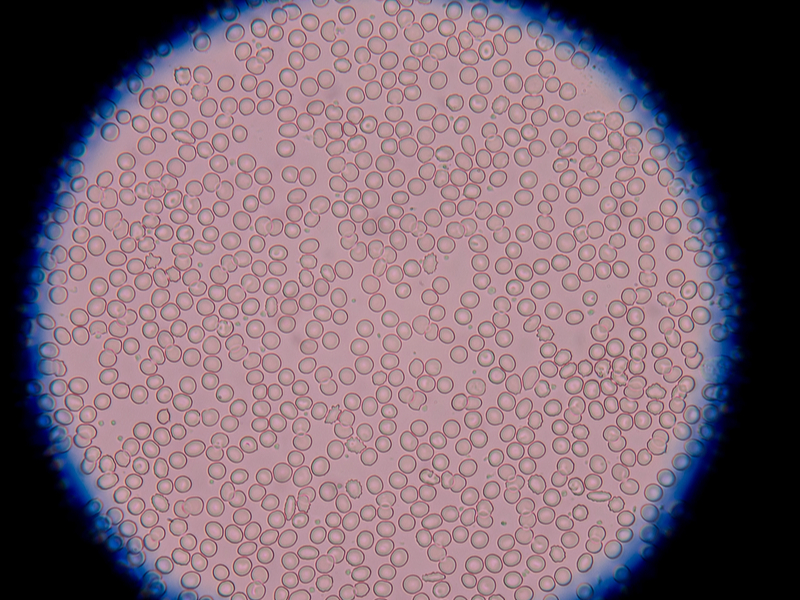
The problem begins when the normal (image left), budding candida species that we all have in our gut undergoes pleomorphic changes and begins forming mycelial or hyphae forms – this form of Candida is pathogenic and can cause a large myriad of symptoms.

This happens when the internal milieu of the gut and other tissues become more acidic, therefore killing off many of the friendly bacteria of the Acidophilus and Bifida species. This can occur either through taking a variety of drugs such as antibiotics, cortisone, anti-inflammatories and chemotherapy used for cancer cases – the bowel flora can also drastically reduce when the pH of the body becomes too acidic – eating sugar and other refined products in large quantities can create this acidity.

It appears that this change in pH can trigger genes in the Candida to begin a pleomorphic change into a stealth organism that is very active – if fed with sugar, it can increase itself from 1 to 100 cells in 24 hours. These 100 cells can then produce 100 each in the next 24 hours, and so on. And so by the 4th day we will have 100 million Candida cells.

Most practitioners trying to treat Canidida will concentrate on killing off the pathogenic, mycelial form, without addressing the significant question of converting this pleomorphic form back to the normal, budding form using the Sanum remedies. Another reason for the large numbers of failures by health practitioners is that the internal milieu is not addressed, and also the diet, which is crucial.  We will come back to these important points when we discuss treatment protocols.

THE PARASITIC ROLE OF CANDIDA

Candida has two parasitic functions:

1. It gobbles up any putrefied food matter in our digestive system (mostly caused by improper digestion due to low stomach acid).
2. After we die, Candida acts to decompose the body by secreting phospholipid enzymes, feeding off our corpse and returning us to Mother Earth. When conditions are right, it transforms itself from the “bud” state into the mycelial state, where filament-like roots invade deep into the mucosa in search of nourishment.

The mycelia release phospholipase, an enzyme that attacks cell membranes of the mucosa, splitting fatty acids, generating free radicals, and causing inflammation in the intestine.

Wherever the yeast colonise they cause symptoms, whether an itchy anus or vagina, diarrhoea, heartburn or sore throat. The mycelial forms release 79 different toxic by-products that damage specific tissues and organs and will determine which symptoms will occur. These mycotoxins can also compete with hormone receptor sites, causing hypothyroidism, hypoestrogenism, as well as binding cortisone, progesterone and other hormones for its own use and causing endocrine deficiency states.

BRIEF HISTORY OF CANDIDA

Hippocrates describes oral candidiasis (around 400 B.C.) as “mouths affected with aphthous ulcerations”. In 1665, Pepys Diary reported “a patient hath a fever, a thrush and a hiccup”, immortalize the idea that oral thrush establish from the host.

In 1771, Rosen von Rosenstein defined an invasive form of thrush.

In 1839, Langenbeck was credited with first recognizing a fungus in a patient with typhoid fever. He describes in detail what is now referred to as septate hyphae, branched pseudohyphae and blastoconidia. However, he recognized the entity to the typhoid bacterium rather than the fungus.

In 1847, French mycologist, Charles Philippe Robin, classified the fungus as Oidium albicans using Albicans to name the fungus causing thrush.

Christine Berkhout and others observed the ability of this fungus to infect humans. Berkhout reclassified it under the current genus Candida, in 1923.  Berkhout’s taxonomy was later made known by the French mycologist, Maurice Langeron and Paul Guerra as “the beginning of the rational systematics of the non- ascosporogenous yeasts”.

OTHER FACTORS CREATING PATHOGENESIS

The pathogenesis of disease associated with Candida in humans is driven by a multitude of factors. Some strains of Candida produce gliotoxin, which may impair neutrophil function. However, Candida is a polyantigenic organism containing up to 178 different antigens, which might explain the number of cross-reactions to yeasts, moulds and even human tissue.

It was shown recently that there is a potential cross reactivity with gluten because of several amino acid sequences that are highly homologous to alpha-gliadin and gamma-gliadin. Such a mechanism might lead to wheat intolerance with its accompanying symptoms, and even trigger Coeliac disease in genetically susceptible people. Furthermore, a placebo-controlled crossover study has revealed that dietary yeast may affect the activity of Crohn’s disease.

Candida produces alcohol and contains glycoproteins, which have the potential to stimulate mast cells to release histamine, and apparently prostaglandin-inflammatory substances that could cause IBS-like symptoms.

MYCOTOXINS

Toxic waste from mycotoxins from Candida infestations can also be absorbed into the bloodstream causing “yeast toxin hypersensitivity”, leading to many symptoms such as anxiety, depression and impaired intellectual functioning.

The main toxin implicated here is acetaldehyde, which is a normal by-product of metabolism, produced in small amounts and rendered harmless by the liver. If however, there is excess production of this by Candida, particularly in low-oxygen environments, and a lack of the appropriate liver enzymes – which tend to be deficient in 5 per cent of the general population – the acetaldehyde will become bound strongly to human tissue. This may cause impaired neuro-transmission in the brain, resulting in anxiety, depression, defective memory and cloudy thinking.

Some 40 – 60% of all immune cells in our body are in the gut. The immune system may also concurrently be adversely affected by poor nutrition, heavy exposure to moulds in the air, as well as an increasing number of chemicals in our food, water and air, including: petrochemicals, formaldehyde, perfumes, cleaning fluids, insecticides, tobacco and other indoor and outdoor pollutants.

Exclusively, yeasts tend to secrete a toxin called gliotoxin, which can disrupt the immune system by inactivating enzyme systems and producing free radicals, thus interfering with the DNA of leukocytes.

The resulting lowered resistance may not only cause an overall sense of ill health, but may also allow for the development of respiratory, digestive and other systemic symptoms. One may also become predisposed to developing sensitivities to foods and chemicals in the environment.

Heavy metals such as mercury and others are found in higher amounts when Candida is present, as the Candida yeasts actually store the metals in their cells – these metals are then released when the Candida die during treatment. This is why it is important to undergo a natural, heavy metals detox while treating Systemic Candidiasis.

Candida is sensitive to a number of antifungal agents, such as Nystatin, which is not absorbed from the gastrointestinal tract after oral administration. It destroys Candida by binding to sterols in the cell membrane, and thereby increasing permeability with loss of cellular contents.

When other health conditions become involved, Candida becomes known as Candida-related complex (CRC). CRC, an excess of Candida in your system can cause a host of uncomfortable signs and symptoms which are syndromes within themselves, such as chronic fatigue syndrome, hypoglycaemia, leaky gut syndrome, fibromyalgia, allergy or sensitivity, hormonal, thyroid, and adrenal dysfunction.

This syndrome isn’t caused by Candida itself present in our mucosal tissues that cause irritation, inflammation, itchiness, redness and pain but by the amount of metabolites from the yeast colonies released inside the human colon when they exceed tolerable amounts.
Patients with CRC often have widespread symptoms affecting multiple organs systems such as:

  • Gastrointestinal symptoms
  • Chronic allergies
  • Unexplained fatigue, always tired
  • CNS fog, mood swings, depression
  • Skin rashes, fungal infections
  • Cravings for sugar, bread, beer

Toxicity in the colon affects the health of the whole body particularly if one’s elimination is slowed as in the case of constipation due to an imbalance in intestinal flora.

The delicate hormonal and chemical balance that orchestrates our emotional health can also be affected causing symptoms of mental illness.

EVERYONE CAN BE AFFECTED WITH CANDIDA!

Anyone can be infected with Candida today! Women can be infected because of antibiotics, steroids, anti-inflammatory medications, hormones and birth-control pills. Men are also being infected with Candida from antibiotics, steroids, anti-inflammatory drugs, pain medications, and sexual relations with an infected partner, even though this often results in a topical infection, not a systemic spread.
Teenagers get Candida from routine treatment with tetracycline or other antibiotics for acne. Babies have Candida from the birth canal or breast milk of the infected mother. That is why babies often have thrush (a white- coated tongue), which is a yeast infection.

Millions of people all over the world are infected with Candida. It is estimated that at least one out of three people in the Western world are affected. Because so many of the population can be infected and because so many factors can cause the condition, Candida is an enormous health problem today and has truly reached pandemic proportions.

Candida coexists in our bodies with many species of bacteria in a competitive balance. Other bacteria act in part to keep Candida growth in check in our body ecology. When health is present, the immune system keeps Candida proliferation under control, but when the immune response is weakened, Candida growth can proceed unchecked. It is an opportunistic organism, one which, when given the opportunity, will attempt to colonize all acidic bodily tissues – this is one of the reasons why it is present in all cancerous tumours as they have a very acidic pH. This uncontrolled growth of Candida is known as “Candida overgrowth” or “Candidiasis”.

UPSETTING THE ECOLOGICAL BALANCE OF THE BODY

Unfortunately, there are many factors in our modern society that can upset the ecological balance of the body, weaken the immune system and thus allow the yeast to overgrow.
The major risks factors are:

  • Steroid hormones, immunosuppressant drugs such as cortisone, which treat severe allergic problems by paralyzing the immune system’s ability to react.
  • Pregnancy and birth control bills which upset the body’s hormonal balance
  • Diets high in carbohydrate and sugar intake, yeast and yeast products, fermented foods
  • Prolonged exposure to environmental moulds
  • Antibiotics and sulpha drugs – probably the chief perpetrator of all-antibiotics that kill ALL bacteria, good and bad. They do not distinguish good bacteria from bad. Antibiotics kill the “good” flora which normally keeps the candida under control. This allows for the unchecked growth of candida in the intestinal tract. It is normally difficult to recover a yeast culture from bodily surfaces.

In a variety of ways the same thing happens with the use of steroid drugs, such as cortisone. All steroids, including the contraceptive pill, will have a depressing effect on the immune system, as well as killing off friendly bacteria in the body.
Yeast infections are common in those that take repeated or prolonged courses of amoxicillin, ampicillin, ceclor, keflex, tetracycline or other broad- spectrum antibiotics during infancy, childhood, adolescence, or since you are become an adult.

DIETARY FACTORS

Sugar is the main nutrient of Candida albicans. Candida albicans thrive on all simple sugars, sweeteners, but also natural sugars in fruits and fruit juices, as well as eating too many complex carbohydrates.
There are several reasons to restrict or eliminate the intake of milk in patients with chronic candidiasis:

  • High lactose content promotes the overgrowth of Candida
  • Milk is one of the most frequent food allergens
  • Milk may contain trace elements of antibiotics, which can further disrupt the gastrointestinal bacterial flora and promote Candida overgrowth

DO YOU HAVE CANDIDA? HOW DO YOU KNOW YOU HAVE IT?

The patient’s history and symptoms is usually the key to arriving at a diagnosis. There are a wide variety of signs and symptoms that are prevalent in systemic Candidiasis, being able to score these systematically provides a good, overall picture. Dr. Crook’s Candida questionnaire is very helpful as it enables the patient to score their symptoms and arrive at a number, anything above 180 for women, and 140 for men is highly significant and represents the majority of symptoms that relate to the Candida.

There are also other testing procedures for diagnosing Candida, using VRT Bioresonance diagnostics such as VEGA Biodermal screening. Using this technology, the practitioner is trying to determine if there is a resonance response with the patient, using an ampoule of pathogenic Candida. If there is no pathogenic Candida in the patient, then there will be no resonant effect, and this is easy to measure using this equipment.

Another method of testing the Candida is to use a form of kinesiological muscle testing called Autonomic Response Testing (ART) invented by a German neurologist Dr. Dietrich Klinghardt, M.D. Ph.D. ART grew out of the importance of detecting and correct problems of the autonomic nervous system (ANS).

ART allows the practitioner to correct the problems of the ANS and to help restore the self regulating mechanism of the body allowing the patient to return to a state of health.

To date, there is no conclusive blood or clinical test for diagnosing pathogenic Systemic Candidiasis. In time to come there will probably be genetic tests that can determine the genome of the mycelial, pathogenic Candida.

Let us now examine the concept of treatment which is quite complex if one really wants to succeed. There are numerous approaches to treating Candida which are used; from medical doctors using medicinal anti-fungals to naturopaths and nutritionists using a variety of natural products to kill off the Candida. All these treatments are rampant with problems that end up in the patient feeling better initially, only to find that the Candida “creeps back” again after a few months.

Dr George John Georgiou, the author of this paper and an internationally renowned Holistic Medicine Practitioner had researched and experimented, treating his own Candida for many years. He spent many years visiting many medical and natural medicine practitioners, but found that no-one was able to treat his Candida successfully and fully. Eventually, after 11 years of trial and error, he managed to put together a protocol that goes further than traditional schools – he has used this treatment protocol for over 10 years now and has successfully treated in excess of 3,500 patients to date.

As this treatment protocol is being implemented at the Da Vinci Holistic Health Center, Larnaca, Cyprus where Dr Georgiou is the Director Founder, it has come to be called the “Da Vinci Candida Treatment Protocol”. Dr. Georgiou now treats patients from all over the world and one of his nicknames is “Dr Candida.”

THE DA VINCI CANDIDA TREATMENT PROTOCOL

There are toxins in the food you eat, the water you drink and the air you breathe. Even your own body produces toxins as a result of its many metabolic processes that keep you alive.

Before implementing the Candida Treatment with patients, Dr Georgiou therefore makes certain that the patient goes through a detoxification protocol to balance the internal milieu. One of the quickest, cheapest and most efficient ways of achieving this is to undergo a 15-day alkaline detox programme using only fresh fruits, vegetables, vegetable juices and soups, steamed vegetables with olive oil and herbs, as well as herbal teas.

There are a number of benefits of detoxification such as:

  • The digestive tract is cleansed of accumulated waste and fermenting bacteria.
  • Liver, kidney and blood purification can take place, which is not possible during regular eating patterns.
  • Mental clarity is enhanced as chemical and food additive overload is reduced.
  • Reduced dependency on habit forming substances such as sugar, caffeine, nicotine, alcohol and drugs,
  • The stomach size is returned to normal as bad eating habits can be stopped.
  • The hormonal system is enhanced which is especially true for growth hormones.
  • The immune system is stimulated.

After detoxifying on an alkaline diet for 15 days, patients report higher energy levels, clear and glowing skin, weight loss of several pounds, clear-headedness, reduced cellulite, good body tone and a great feeling of being relaxed.

THE DA VINCI CENTRE DETOXIFICATION DIET

We suggest that the person eat only fresh fruit, salads, freshly squeezed juices, steamed vegetables and vegetable soups for 15 consecutive days.

These are the foods that are allowed during the detoxification phase, no other family of foods is allowed. You may eat as many of the following foods as you wish, but it is best to eat only when you feel hungry. Wash all fruit and vegetables in a bowl of water with 4-5 tablespoons of grape vinegar added to help wash away any pesticide/ herbicide residues. Rinse afterwards with clean water.  Here are the foods that you can eat in plenty:

Salads – use any type of fresh vegetables you like, in any combination. Use organic vegetables when available, and include bean sprouts when in season. Salad dressings should be kept simple- a little virgin olive oil with fresh lemon, or cider vinegar. Add plenty of fresh onion and garlic- these are very detoxifying.

Steamed vegetables – eat any variety you like, including broccoli, cauliflower, potatoes, beetroot, carrots etc. Steam as opposed to boil, and eat with a little herbal salt, lemon and a little virgin olive oil, with plenty of garlic.

Vegetable/fruit juices – drink a minimum of 1-3 per day, and try to include one cocktail comprising one- third of a glass of raw green juice (spinach, parsley, cabbage and any other green vegetables), topped up with carrot juice. Carrot juice has a strong effect on the digestive system, provides energy, serves as an important source of minerals, promotes normal elimination, has diuretic properties and helps to build healthy tissue, skin and teeth.

Fresh Fruit – choose the fruit of your choice and eat as much as you like, whenever you like. You could begin the day with 2-3 pieces of fruit, which are gentle on the digestive system. Make a tasty fruit salad. Try to avoid too many juicy fruits as this may overly feed the Candida, and certainly avoid all forms of fruit juices as this will give sugar to the body quickly, again feeding the Candida. There is tremendous benefit in adding fruit during the detox due to their living enzymes and phytonutrients which are very cleansing to the body. Remember, we are not treating the Candida yet, only helping the body to cleanse and prepare for the 3-month Candida protocol.

Herbal teas – choice any of your choice. Chamomile is a good relaxant, aniseed and mint is good for the digestive system, Kombucha, dandelion tea which is excellent for purifying the blood and detoxifying and stimulating liver function, sage tea which is a blood cleanser, nettle tea which is excellent for driving away excess fluid out of the tissues and is a wonderful cleanser for all the detoxification organs. Drink as many as you like, with a little honey on the tip of a teaspoon if you like.

The purpose of this diet is to detoxify – to remove the toxins from the fat cells and tissues as well as the organs, so that the body can return to its optimum level of functionally.

PARASITES, HEAVY METALS AND OTHER TOXINS

As part of the detoxification process, the Da Vinci Center also attempts to detoxify heavy metals that can easily be detected using a hair analysis sent to a US laboratory of repute.
In addition, there are also parasites which more than 90% of people carry, so it is good to also incorporate a parasite cleanse in with the 15-day detox diet.

Heavy metals are found from fish and amalgam fillings (mercury): aluminium found in cheeses, baking powders, cake mixes, self- raising flour, cosmetics, toothpastes, antiperspirants and some drugs such as antacids.

Arsenic is given to chickens as a growth-promoter, cadmium is found in tea and coffee, as well as cigarette smokers. Lead is found in paints, fuels, rubber, plastics, inks, dyes, toys, building materials and hair restorers.

One of the most researched natural chelators in the world that has undergone double-blind, placebo controlled trials with 350 people is called HMD™. It has been shown to safely chelate many different types of heavy metals, including uranium, a difficult metal to chelate.

As part of a novel approach to eradicating parasites, the use of small, portable bioresonance devices invented by Russian scientists called Deta Elis have been used with good success.

Let us now examine the Da Vinci Candida Treatment Protocol in more depth.
The Da Vinci Candida Protocol has five basic objectives:

  1. Starve the Candida by eliminating the foods that feed it
  2. Kill the Candida through the use of anti-Candida natural products
  3. Repopulate bowel flora with a high-potency probiotics such as Custom Probiotics, an excellent American company that specializes only in probiotics – the capsules are guaranteed to contain 60 billion live bacteria one year after manufacture.
  4. Regulate the dysbiosis and convert the pathological, mycelia form of Candida back to the normal form by the use of the Sanum remedies produced by the Sanum Kehlbeck company in Germany.
  5. Restore biochemical balance to the body and strength to the immune system. This will allow the body once again to regain and maintain control over Candida growth by optimizing the diet – this would involve avoiding food intolerances and following the Metabolic Type diet by Bill Wolcott. Also kill off parasites and chelate heavy metals out of the system.

Let us examine these steps in a little more details below:
Phase 1 – Starving the Candida
The foods that should be strictly AVOIDED during that time include:

  • Sugar– and all foods that contain sugar. These include white and brown sugar, honey, syrups, liquor, lactose, fructose, all confectionary and sweet cakes, chocolates, ice-creams, home- made sweets and cakes, biscuits, fizzy beverages and all fruit drinks.
  • Yeasts – and all foods that contain yeast including breads, vinegar, ketchups, mayonnaise and pickles.
  • Mushrooms– all types, including Chinese mushrooms such as Shitake
  • Refined foods– all white flours, white rice, white pasta products, corn flour, custard and refined cereal products, unless they are whole meal or organic.
  • Fermented products – all alcoholic beverages, vinegar and all vinegar products such as ketchup, mayonnaise and pickles, beer.
  • Nuts – all types of nuts that are cleaned and packaged without their shells- these have a tendency to collect fungal spores and moulds from the atmosphere, which will antagonize the Candida. Nuts that are fresh with their shells are OK.
  • Fresh and dried fruit – all fresh fruit should be avoided for the six weeks only as again, the fructose they contain will feed the Candida and make it extremely difficult to eliminate.

All other fruit that is not fresh such as cooked, tinned or dried and fruit juices should be avoided for the full 3 months.

Phase 2 – Killing the Candida
There are a number of herbal formulas, homoeopathic, isopathic remedies (Sanum) and probiotics that are used in the Da Vinci Candida Protocol – they have been carefully selected after years of experimentation, and the fact that they have worked time and time again with hundreds of people. The aim of using these supplements is to kill off the Candida.
Here are the supplements in order

  • Kandidaplex– a doctor- formulated compound that contains Berberine, undecylenic acid, biotin, Sorbic acid, Pau D’ Arco and resveratrol from Japanese knotweed (Polygonum cuspidatum) root extract.

KandidaPlex™ features calcium undecylenate, a fatty acid that helps the body balance colonization of Candida. This formula also includes natural ingredients such as pau d’arco, berberine, and a vegetarian enzyme blend, formulated specifically to balance the growth of Candida albicans and improve the intestinal microenvironment.*Dosage: 2 capsules x 3 daily

  • Horopito – a New Zealand herbal product that contains two powerful anti- fungal agents that have been shown to kill Candida- Pseudowinterata Colorata and the synergistic herb aniseed, that boosts effectiveness 6 fold. This herb has undergone scientific research and it has been found to be as effective as Nysteral and other anti-fungal medications, but without the side effects. Dosage: 1 capsule twice daily
  • Caprylic acid (600 mg) – a derivative of coconut that stops the Candida reproducing, as well as killing the Candida.Dosage: 1 tablet x 3 daily
  • Candida 30c homoeopathic nosode made from pathogenic Candida to kill off the Candida homeopathically.Dosage: 2 pillules x 3 daily for 2 weeks only. These are stopped just as the Sanum remedies are begun.

Phase 3 – Repopulating the Friendly Bacteria
This phase runs parallel with phase 2 and uses good quality, human strain probiotics such as the high-potency Custom Probiotics containing five superior strains of freeze-dried probiotic microorganisms that specifically target and help the small and large intestines, improving the intestinal microflora and digestive processes. These are:
• L. Acidophilus
• L. Rhamnosus
• L. Plantarum
• B. Lactis
• B. Bifidum

At the time of manufacture, each capsule actually contains 96 billion live bacteria, but are guaranteed to have a minimum of 60 billion one year later. They are also resistant to higher temperatures for a short period of time, making them convenient for travel for small periods of 2-3 weeks. Dosage: 1 capsule x 3 daily

To these supplements we add a good- quality multivitamin such as HMD MULTIS (2 caps x 2 daily) as well as Omega 3,6,9 fatty acids (1 caps x 2 daily) to provide all the vitamins and minerals that the immune system requires for optimal functioning.

Phase 4 – Using Isopathic Remedies to Normalize Pathogenic Candida 
All the above must be taken for the full 3 calendar months of the protocol, with the exception of the homeopathic Candida 30c. After two weeks of the anti-Candida diet, certain specialized isopathic remedies are introduced, known as Sanum remedies from Germany, after the work of Prof. Enderlein, the father of pleomorphism and live blood analysis. Each of these isopathic remedies is only taken a couple of times per week, as follows – see also table below:

Mucokehl D5 tabs – 1 tab, twice weekly
Pefrakehl D4 caps – 1 cap twice weekly
Notakehl D5 tabs – 1 tab twice weekly
Nigersan D5 tabs – 1 tab twice weekly
Albicansan D4 caps – 1 cap every second day

Table showing schedule for taking Sanum remedies
If there is vaginal discharge, or anal Candida, then vaginal or anal pessaries of Albicansan D3 must also be used to eliminate this topical infection. These can be used every second day last thing at night, after sex.

These Sanum remedies mentioned above are continued for 10 weeks until the end of the Candida protocol. It is wise to begin the Sanum remedies TWO WEEKS after beginning the general protocol in order to allow a considerable portion of the Candida to die off, and to reduce the severity of the Herxheimer reaction.

Fruit can be re-introduced back into the diet FOUR WEEKS after the beginning of the Sanum remedies.

All capsules and tablets should be taken away from food and should not be taken together, as they clash. Follow this simple table below of how and when to take your Sanum remedies. These remedies are taken BEFORE or AWAY from food.

Open the capsules and pour the powder that they contain under the tongue and allow it to dissolve and absorb for a few minutes.
Prof. Enderlein’s Sanum remedies work by changing the harmful microorganisms in the body fluids into non- aggressive forms, probably by changing the pH and electrical conductivity. Harmful bacteria and toxins are broken down and excreted through natural processes. They also help to alleviate the dysbiosis and bring the internal mileau of the intestine back into balance.

Phase 5 – Balancing Body Chemistry    
It is a commonly recognized and accepted fact that immune system efficiency is highly dependent on the proper biochemical balance in the body. This depends on proper and adequate nutrition to supply the body with all the required biochemical constituents (vitamins, minerals, enzymes, intrinsic factors).

Different people require different amounts and balances of nutrients for optimum health. The criteria for the determination of these differing nutritional requirements lies within the definition of one’s metabolic type, i.e., the genetically determined metabolic and nutritional parameters that define each person’s individuality on every level.

It is precisely because different people have different metabolic types, and therefore different needs for nutrition, that the allopathic, symptom- treatment approach in nutrition is baseless and so often ineffective.

The Blocking Factors of Recovery
Many time people who have Candidiasis don’t follow the Candida protocol precisely, consistently, and for a long enough period of time. Often when people feel better while they are on the Candida diet, they tend to go off it too quickly.

Often they don’t understand the need for its limitations, the importance of staying in tract, or the many substitutions that are available for problem foods or ingredients.

It has been found and research that it is beneficial to stay on the protocol until optimum health has been reached to the levels of treated.
Many people make the mistake of not rotate their foods on the protocol, and they repeat the same food more often. If they have a food family too often, they could easily develop intolerance to it, because the digestive system of the Candida sufferer is generally quite sensitive.

When people have Candida or CRC they usually have weakened immune systems. When the immune system is suppressed, it is prone to more infections and disease, making it impossible for a person to fully recover.

Depending on the severity of Candida overgrowth and the amount of the agents taken, the Candida can be killed off in vast numbers in a very short period of time. As they are killed, they release substances that are toxic to the body- mycotoxins. If the elimination organs such as the kidneys, liver, lymphatics, gut and skin cannot clear these mycotoxins quickly and then accumulate in the tissues, then a temporary toxic or allergic-type reaction can occur. The technical name for this experience is a Herxheimer reaction but it is more commonly referred to as “die off”.

Usually “die off” lasts only a few hours, though it can last several days. It can usually be controlled by reducing the dosage of the remedies used to kill the Candida, as well as taking drainage herbs and homoeopathics that your practitioner will advise you on.

Signs of Herxheimer reaction can be many and varied bur generally involve such discomfort as aching, bloating, dizziness, nausea.  Exercise as well as insuring proper, daily bowel evacuation has been reported as being helpful in countering the adversities of die- off. Maintaining a high daily intake of pure water is also important to keep the channels of elimination open. Sometimes taking a teaspoon of baking soda (sodium bicarbonate) in a glass of water can help to quickly neutralize acidic reactions in the body that lead to inflammation and pain.

It may be possible to slow down these symptoms, many of which are caused by acetaldehyde, one of the main toxins produced by yeast. Taking molybdenum can break down this toxin into something far less harmful.

Adrenal gland dysfunction can create a complication to Candida recovery. This will affect the person’s ability to feel energized, cause the person to feel hot or cold temperatures more easily, possible sweat more than usual, heart palpitations, low sugar levels.

Lack of Hydrochloric acid (HCL)
HCL is produced in the stomach to aid in activating digestion of foods and protection of the intestinal flora.
One of the most common causes of HCL imbalances is past antibiotic use. Antibiotics destroy the beneficial bacteria that synthesize B vitamins necessary for HCL production in the stomach. Proper HCL levels in the stomach kill off many pathogens that otherwise would enter into the intestinal tract and potentially create problems.

HCL’s important functions include:

  • Breaking down proteins into essential amino acids and nutrients
  • Stimulating your pancreas and small intestines to produce the digestive enzymes and bile necessary to further breakdown the carbohydrates, proteins and fats
  • Preventing disease by killing pathogenic bacteria and yeast normally present in foods.
  • Low stomach acid leads to a cascade of digestive problems such as bloating, gas and constipation.
  • Preventing the return of Candida

So if the patient suffers from hypochlorhydria or low production of hydrochloric acid from the stomach, then add a Betaine HCl + Pepsin supplement. In addition, if there is bloating, flatulence and a general discomfort of the gut, then one can add pancreatic enzymes too.

Natural Antibiotics During Candida Protocol
It is critical that anyone on the 3-month Candida protocol stock-up on NATURAL ANTIBIOTICS that they might require if they come down with a cold, flu, sore throat or any kind of infection while they are still on the Candida protocol.

These natural antibiotics have been tried and tested for many years and seem to work fine with most infections. However, it is very important to take these immediately when the first symptoms appear. If you leave the infection for a couple of days the microbes will spread quickly and it will be more difficult to shift it with the natural antibiotics.

This is why it is critical to have these natural antibiotics in your medical dispensary BEFORE beginning the Candida protocol – they have an expiry date of between 2 – 5 years, so you will no doubt use them during this time.

The dosages mentioned below are for adults. For maximal effectiveness it is good to use at least 4 of the natural antibiotics mentioned below, all in combination together.

These Natural antibiotics and herbal supplements include – link all the yellow to the site:

  • Grapefruit seed extract (Citricidal):  is a very effective anti-fungal, available in tablet form as well as liquid form which can be placed under nails with fungus. Take two tabs x 3 daily. For nails, one drop under each nail morning and evening.
  • Oregano Oil – if one is citrus intolerant, then you can use Oregano oil gel caps instead – one gel cap x 3 times daily.
  • Silver Liquid 50 ppm – you can take one teaspoon x 3 times daily.
  • Vitamin C – take either 2 tabs (1000mg) x 3 daily, or ½ tsp x 3 times daily of the calcium ascorbate powder form.
  • TRIFORM – a combination of a number of antibiotic herbs such as poke root, Echinacea, Berberine and more. Take 35 drops x 3 daily.
  • Olive Leaf Extract – good to take one cap x 3 times daily if you know it is a viral infection.

If you regularly suffer from colds and flus, then it would be good to take about 1/3 tsp of vitamin C powder x 3 times daily – take this throughout the Candida Protocol. In addition, one can also take an immune-modulating product called EPICOR – one cap x 2 times daily.
Over the years Dr Georgiou has successfully treated in excess of 3,500 patients to date – here are just a few cases as examples:

CASE HISTORIES

CASE NO 1

Mrs. A, Age 44
Mrs. A’s presenting symptoms were somewhat unusual in that she continually complained that she frequently had the sense of a strong fishy odour in her nostrils over the last 7 years.

She remembers that this began when she had cleaned mould in her house with chlorine – the mould had appeared after a flood.
She also suffered from many allergies which included from  a number of flowers, bananas and melon.

Her main symptoms apart from the annoying fishy odour was, constant intermittent coughing as well as a heavy pressure-type sensation in the chest and lungs. She had clear signs of nail fungus as well as frequent vaginal discharge.

She had consulted a number of medical doctors and dermatologists, but with no success. The dermatologist gave her antifungal cream for the nails. The condition remained as before.

She underwent bioresonance diagnosis using the VEGA system and was found to have a number of food intolerances too such as: wheat, lactose and milk products, bananas, caffeine, sugar, chicken, pork, nightshade family of vegetables (potatoes, tomatoes, peppers, and aubergines), olive oil and olives.

The VEGA test also showed that she was resonating with pathological, mycelial forms of Candida albicans, indicating that she was suffering from systemic Candidiasis – mixed moulds were also found during this testing protocol.

It was decided to help her body to detox and return back to an alkaline pH, as well as help eliminate inflammatory chemicals and other toxins. She followed an alkaline detoxification diet for 2 weeks based on alkaline foods such as fruit and vegetables.

Her energy levels after the detox had tremendously increased and she reported clarity of mind. She began the Da Vinci Candida Protocol for 3 months – see main text for details.

The smell of fish had decreased by 30% in intensity and the frequency to cough had also decreased by 40% within the first 3 weeks of the protocol treatment. After two months of treatment, the cough had improved 60%. Previously she would cough for one hour, now she coughs less than one minute. The smell of fish has improved by 70%.

After the completion of the Candida protocol (3 months), the Candida finally disappeared and the cough had improved by 100%. This was the first time that the cough had improved in the past 7 years. The smell of fish had also vanished as well as the nail fungus on her toes – this was also helped by adding grapefruit seed extract liquid – one drop per nail morning and evening. She was a very happy woman!

CASE NO 2

Mr. M, Age 45
Mr. M presented with a chronic cough that he had for the past 7-8 years, accompanied by whitish phlegm. He was diagnosed with H. Pylori for which the medical doctor prescribed antibiotics which were taken on and off for a period of 2 years. The coughing however persisted even though he had consulted many doctors, including ENT (otolaryngologists) and pneumonologists with no results. Before coughing began he lived in a mouldy apartment.

He underwent VEGA food intolerance testing that showed  intolerance to a number of foods such as: wheat, soya, lactose and milk products, beans, caffeine, almonds and walnuts, pork, citrus (lemon, grapefruit, and oranges), olive oil and olives. He is a vegetarian but includes lactose and fish in his diet.

The VEGA testing also showed that he was suffering from systemic Candida albicans.
He followed an alkaline detoxification diet for 15 days based on fruit and vegetables. During the detox the cough had decreased by 50% in frequency and 70% decrease in intensity. The white phlegm had stopped completely.

Immediately after the detox he began the Da Vinci Candida protocol for 3 months. During the Candida protocol his energy levels had massively increased and he had incredible clarity of mind. After the Candida treatment his cough had completely vanished and the phlegm decreased to minimum.

CASE NO 3

Mrs. S, Age 49
Mrs. S complained that she had bronchial asthma and suffered from allergies. Medical doctors gave her cortisone sprays. She also suffers from obesity (147 kg) and whenever she tries to diet she suffers from hypoglycaemia.

Other health issues include atrial fibrillation (cardiac arrhythmia) and GERD- gastroesophageal reflux disease. She was taking Warfarin, an anticoagulant, to prevent blood clots from forming. She was also taking  medication to control her arrhythmia.
She had also removed her thyroid nodules and was taking thyroxine daily.

It was recommended that she begin with a compromised alkaline detoxification diet lasting 1 month. This means that the body will be detoxing slower than the 15 days alkaline detox diet by leaving in protein foods the first two weeks. This procedure is recommended when there are chronic degenerative disease in order to prevent any possible adverse reactions caused by the elimination of inflammatory chemicals.

So, the first week of detox she was allowed to eat fish and pulses along with fruit and vegetables. The second week the fish was eliminated and only the pulses remained along with the fruit and vegetables. During the final two weeks she only ate fruit and vegetables.

During the detox her stomach digestion improved and she was feeling much better; oedema had also disappeared after some heavy urination initially, and she had lost noticeable inches around her waist – to her delight as weight had been blocked for a long time.

During bioresonance testing it was shown that she was intolerant to a number of foods such as: wheat, lactose and milk products, citrus (oranges, lemons, grapefruits), caffeine, sugar, hazelnut, walnuts, almonds, pork, chicken, and nightshade family of vegetables (potatoes, tomatoes, aubergines, peppers).

In addition, the Vega Bio-dermal testing also showed that she was suffering from systemic Candidiasis. She therefore began the Da Vinci Candida protocol for 3 months. During the Candida protocol she had lost a total of 12 kg, her asthmatic symptoms had gone completely, and she could now climb steps without wheezing and panting, being much quicker on her feet. As an added gift her chronic sinusitis had also completely cleared.

CASE NO. 4

Mrs J, 70 years old
We will be brief here – a 70 year old lady who had psoriasis most of her life was treated for systemic Candidiasis using the abovementioned protocol and within 90 days the psoriasis had cleared.
Before beginning the Candida protocol

3-months after completing the protocol


REFERENCES
Truss, CO. Metabolic abnormalities in patients with chronic Candidiasis – the acetaldehyde hypothesis, Journal of Orthomolecular Medicine, 13:63- 93, 1984
Gail Burton. Candida – The Silent Epidemic, Candida Causative factors, 4-9, 2003
Leon Chaitow N.D, D.O, Candida Albicans- Could yeast be your problem? How Candida gets out of hand. Chapter 3- Immune System Deficiency, 24-26, 1991
John Parks Trowbridge, M.D., and Morton Walker, D.P.M., The yeast syndrome- Antibiotics Encourage Yeast Overgrowth, 45-46, 1986
William G. Crook, M.D., Chronic Fatigue Syndrome and the yeast connection, Probiotics: 260,261, 1992
William G. Crook, M.D., The yeast connection – Candida questionnaire and score sheet – Diagnosis of a yeast-Related disorder, 29-33, Food allergies: 122, 1986
Hulda Regehr Clark, Ph.D., N.D., The Cure for all Diseases 1995
Crandall M. The pathogenetic significance of Intestinal Candida Colonization, 2004
Budtz- Jorgensen, E. Cellular immunity in acquired Candidiasis of the palate. Scand. J. Dent. Res. 81, 372, 1973
Hussein, H.S., and J.M. Brasel. Toxicity, metabolism, and impact of mycotoxins on humans and animals. Toxicology 167, 2001
Calderone, R.A., and R.L. Cihlar (e.d.). Fungal pathogenesis: principles and clinical applications. Marcel Dekker, Inc., New York, N.Y, 2002
Trowbridge JP and Walker M. The yeast Syndrome Bantam Books, New York, N.Y, 1986
Truss C: The missing Diagnosis Birmingham Alabama (The Author), 1983
Crook WG: The yeast connection, A medical Breakthrough 2nd Addition Professional Books, Jackson, TN, 1984
Michael T. Murray, N.D., Chronic Candidiasis, Dietary factors, sugar and the yeast syndrome: 43,44, 1997
Dr Georgiou has been an active clinician for 30 years, and Director Founder of the Da Vinci Holistic Health Centre and the Da Vinci College of Holistic Medicine in Larnaca, Cyprus